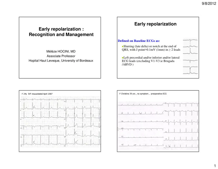
early repolarization early repolarization recognition and
play

Early repolarization Early repolarization : Recognition and - PowerPoint PPT Presentation

9/8/2012 Early repolarization Early repolarization : Recognition and Management Defined on Baseline ECGs as: Slurring (late delta) or notch at the end of QRS, with J point>0.1mV (1mm) in 2 leads Mlze HOCINI, MD Associate


  1. 9/8/2012 Early repolarization Early repolarization : Recognition and Management Defined on Baseline ECGs as: � Slurring (late delta) or notch at the end of QRS, with J point>0.1mV (1mm) in ≥ 2 leads Mélèze HOCINI, MD Associate Professor � Left precordial and/or inferior and/or lateral Hopital Haut Leveque, University of Bordeaux ECG leads (excluding V1-V3 ie Brugada /ARVD ) F Christine 39 yrs , no symptom , preoperative ECG F 24y IVF resuscitated April 2007 1

  2. 9/8/2012 F Christine 1yr later: Sudden cardiac arrest while working on computer Evidence of the pathological relationship with SCD 2

  3. 9/8/2012 type of « Early Repolarization » Evidences of the pathological relationship with SCD up to 50% of highly Malignant form trained athletes 1- Prevalence of « early repolarisation » - 31% ( 64/206) in pts with IVF vs 5% in the matched group (412) (p=0.002) (Haissaguerre NEJM 2008) - 60% of 15 patients with IVF (vs 3.3% controls ) (Nam, NEJM 2008) - 42% in pts with IVF vs 13% in young athletes (Rosso et al. JACC 08) - 5,8% in the general population ( Tikkanen JT et al. NEJM 2009) 2- Amplitude of J point - 2.15±1.2mm in IVF vs 1.05±0.2mm in controls with « early repolarisation » Corrado et al . European Heart Journal (2010) 31, 243–259 Evidences of the pathological relationship with SCD M 15y M 17y M 45y M 38y I I I I 1- Prevalence of « early repolarisation » II II II - 31% ( 64/206) in pts with IVF vs 5% in the matched group (412) (p=0.002) (Haissaguerre NEJM III II III 2008) aVF - 42% in pts with IVF vs 13% in young athletes (Rosso et al. JACC 08) III aVF - 5,8% in the general population ( Tikkanen JT et al. NEJM 2009) V1 aVF III V1 2- Amplitude of J point V1 aVF repolarisation » - 2.15±1.2mm in IVF vs 1.05±0.2mm in controls with « early V1 V4 V4 3- Dynamicity of J wave: Instantaneous J/ST changes, V4 Accentuation of repolarisation at the time of Arrhythmias V4 V5 J/ST elevation from 2.6±0.1 to 4.1±2mV (p<0.001) V5 V5 V5 ECG of VF initiation in 18 pts V6 V6 V6 V6 Inferior 44% Lateral 9% Inferolateral 47% 3

  4. 9/8/2012 Accentuation of repolarisation at the time of Arrhythmias Instantaneous J/ST changes M 52y valsalva * * * * * * F 16 y night * * * * * Evidences of the pathological M 52y Familial nocturnal SD ECG minutes after admission 5d later 0.1mV 0.2mV relationship with SCD I I 1- Prevalence of « early repolarisation » II - 31% (64/206) in pts with IVF vs 5% in the matched group (412) (p=0.002) (Haissaguerre NEJM 2008) II - 42% in pts with IVF vs 13% in young athletes (Rosso et al. JACC 08) III III - 5,8% in the general population ( Tikkanen JT et al. NEJM 2009) aVR 2- Amplitude of J point aVR - 2.15±1.2mm in IVF vs 1.05±0.2mm in controls with « early repolarisation » aVL aVL 3- Dynamicity of J wave: Accentuation of repolarisation at the time of Arrhythmias - J/ST elevation from 2.6±0.1 to 4.1±2mV (p<0.001) ECG of VF initiation in 18 pts aVF aVF 4- Correlation location J/ST and Arrhythmia origin V1 V1 Most VPB positive in V1–V2 (LV origin) Endocardial mapping and V2 ablation in 8 pts V2 V3 ST elevation localized in inferior leads associated with superior axis V3 (origin in the inferior wall) V4 V4 Widespread abnormal repolarization associated with extreme V5 polymorphism in other ST location V5 V6 V6 500msec 500msec 4

  5. 9/8/2012 Two ECGs 24 hours apart Results : Clinical data I 64 patients EAR ; 10 had a familial history II of SCD 18 female , 46 male, 35±13 years III – VF occurred during normal physical activity in 26, at aVR rest in 15, sleeping in 12* and effort in 6 – Preceding syncope in 24 (37%) : prior ECG available aVL described as ‘borderline or normal variant or early repolarization’ aVF M 22yrs Beat to beat fluctuations favor Repolarization rather than Depolarization V 1 D I D I V 1 * * I I V 2 D II D II V 2 II II V 3 D III III D III V 3 aVR V 4 aVR III aVR V 4 aVL aVL V 5 aVR aVL V 5 aVF V1 aVF V 6 aVF V 6 aVL V2 April 2004 March 2006 aVF V1 5

  6. 9/8/2012 FOLLOW-UP 60 ±42 months H 34yr, convulsions while sleeping, 2 ECGs the same day Probability of no recurrence 7h 1,0 0,9 0,8 IVF:23% recur 0,7 0,6 0,5 0,4 0,3 IVF+ Repolarization 0,2 Abnormality: 43% recur 0,1 15h 0,0 0 1 2 3 4 5 6 7 8 9 10 Time (Years) No early repolarization Early repolarization Resistance to AA drugs of class Ib (7/7), Ic (10/10), BB (11/11), Amiodarone (7/8) N Engl J Med. 2009 Nov 16. 10864 patients (44 ±8 yo) Follow-up 30 ±11 years RISK STRATIFICATION • J-point elevation in inferior leads – >0.1mV (5.8%) � RR cardiac death: 1.30 (CI:1.05 – 1.61, p=0.02) – >0.2 mV (0,33%) � RR cardiac death: 3.03 (CI 1.88- 4.90, p=0.001) � RR arrhythmic death: 2.99 (CI 1.49-6.03, p=0.005) Stronger predictors than QT interval and LVH 6

  7. 9/8/2012 Horizontal/descending ST segment Provocative manoeuvers in Early Repolarization Ascending ST Horizontal/descen • 206 patients with IVF included segment ding ST segment • 142 without Early Repolarization � benign � poorer outcome • 64 with Early Repolarization outcome ERS No ERS Risk of cardiovascular mortality HR: 8.75 (CI 3.48- 22.0, p<0.0001 From different centers 31 non inducible France 10/28 (Bordeaux 6/10) � Germany 1/7 � Belgium 3/4 � Inducibility per center: 16 inducible Japan 2/3 � From 0% to 75% Swiss 0/3 � Finland 0/2 � EPS n=47 of 64 Tikkanen J et al. Circulation 2011 Rosso R et al Heart Rhythm 2011 Haïssaguerre et al. nejm 2008 Use of Body Surface Mapping Substrate of Ventricular Fibrillation with early repolarization Twin brother H 19y Male 23 years Resuscitated DI aVr V1 V4 DI aVr V1 V4 SD/ VF while walking H 19 y, his twin brother died from DII aVL V2 V5 DII aVL V2 V5 unexplained sudden death : RV evidence for inhomogeneous area DIII aVF V3 V6 DIII aVF V3 V6 LV inferior Y Rudy et al 7

  8. 9/8/2012 No Pharmacological test to depict malignant Early Repolarization 27pts 40 IVF/70 controls Experimental background: Antzelevitch work Yan, G.-X. et al. Circ 1996 – No change : Ajmaline, flecainide, cibenzoline, 1- pre- and post–J- pilsicainide, verapamil , epinephrine, ATP, Ca wave amplitudes were larger with pause- – Slight accentuation : bradycardia, Betablockers dependent 2- augmentation only – Decrease: in pts with IVF • with Exercise/Isoproterenol (7/7pts) (increase in ICa- (specificity and ppv L current thus decrease electrical gradient, increase 100%) HR and reducing inactivation of Ito • and under Quinidine* (9/9pts) inhibits Ito. • Both are powerful treatments for arrhythmic storms or multiple VF. Haissaguerre et al, JACC 2009 Aizawa et al JACC 2012 Multiple episodes of VF and immediate correction by Iso infusion Multiple episodes of VF, correction by Quinidine 2010 2002 2007: 5 years later ICD shocks 14 yo girl No recurrence under quinidine Blood level 1.1 µg/ml with > 50 ICD shocks bisulf. 600mg bid I II III aVR aVL aVF V1 V2 V3 V4 V5 V6 Bernard A et al. JICE 2009 8

  9. 9/8/2012 10 yo girl with syncope One month later Take Home Messages Brother died suddenly at age17 600 mg Hydroxyquinidine/day • In the setting of resuscitated SCA • ICD ±Isoproterenol in case of arrrhythmic storm ±Quinidine in case of recurrence • Familial screening Take Home Messages Take Home Messages • In the setting of resuscitated SCA • In the setting of resuscitated SCA • ICD • ICD ±Isoproterenol in case of arrrhythmic storm ±Isoproterenol in case of arrrhythmic storm ±Quinidine in case of recurrence ±Quinidine in case of recurrence • Familial screening • Familial screening • In the setting of syncope • In the setting of syncope • Characteristics of the syncope • Characteristics of the syncope • Familial history of SCD • Familial history of SCD > Clinical follow-up, ILR, ICD > Clinical follow-up, ILR, ICD • In asymptomatic patients • No pharmacological test yet • EPS seems useless for risk stratification • Useful for screening in familial SCD? 9

  10. 9/8/2012 Conclusions • The 2 major risk factors for VF in the setting of J wave elevation in infero-lateral leads are syncope and major J wave elevation . • Isoproterenol and quinidine decrease the J wave amplitude and prevent VF recurrence. • At present, we are lacking a pharmacological test to screen asymptomatic patients at risk of SCD. 10

Download Presentation
Download Policy: The content available on the website is offered to you 'AS IS' for your personal information and use only. It cannot be commercialized, licensed, or distributed on other websites without prior consent from the author. To download a presentation, simply click this link. If you encounter any difficulties during the download process, it's possible that the publisher has removed the file from their server.

Recommend


More recommend