Disclosure UCSF Current Issues in Pathology 2019 Company - - PDF document

disclosure
SMART_READER_LITE
LIVE PREVIEW

Disclosure UCSF Current Issues in Pathology 2019 Company - - PDF document

5/25/19 Disclosure UCSF Current Issues in Pathology 2019 Company Relationship type Presage Biosciences Consultant A practical approach to well- differentiated adipocytic tumors Andrew Horvai MD PhD Clinical Professor, Pathology UCSF, San


slide-1
SLIDE 1

5/25/19 1

A practical approach to well- differentiated adipocytic tumors

UCSF Current Issues in Pathology 2019

Andrew Horvai MD PhD Clinical Professor, Pathology UCSF, San Francisco, CA

Disclosure

Company Relationship type Presage Biosciences Consultant

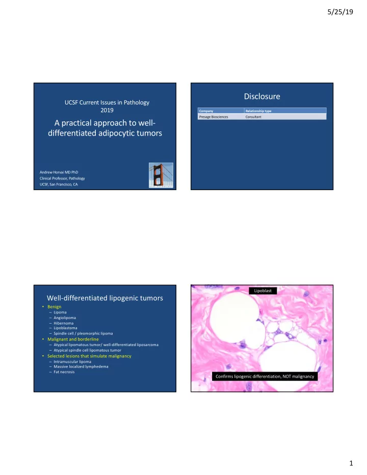
Well-differentiated lipogenic tumors

  • Benign

– Lipoma – Angiolipoma – Hibernoma – Lipoblastoma – Spindle cell / pleomorphic lipoma

  • Malignant and borderline

– Atypical lipomatous tumor/ well-differentiated liposarcoma – Atypical spindle cell lipomatous tumor

  • Selected lesions that simulate malignancy

– Intramuscular lipoma – Massive localized lymphedema – Fat necrosis

Lipoblast Confirms lipogenic differentiation, NOT malignancy

slide-2
SLIDE 2

5/25/19 2

64 year old man, thigh mass Pedunculated thigh mass Lipoma (conventional)

  • Most common soft tissue neoplasm of adults
  • Painless slow growing subcutaneous mass
  • Uniformly benign, no recurrence
  • Circumscribed, unencapsulated
  • Mature adipose tissue, uniform cell size
  • No atypia
  • Ancillary tests: S100+
  • Variants

– Intramuscular, lipomatosis, with metaplastic cartilage CLINICAL PATHOLOGY VARIANTS

45 year old woman, painful forearm mass

slide-3
SLIDE 3

5/25/19 3

Angiolipoma

  • Subcutis of forearm, multifocal, painful, multifocal
  • Familial forms
  • Uniformly benign, no recurrence
  • Circumscribed, thinly encapsulated
  • Mature adipose tissue, peripheral or diffuse capillaries

with fibrin

  • No atypia
  • Ancillary tests: PTAH
  • Variants

– Cellular angiolipoma Angiolipoma: PTAH Cellular angiolipoma

CT Axial PET

25 year old woman with right groin mass

slide-4
SLIDE 4

5/25/19 4 25 year old woman with right groin mass

Hibernoma

  • Rare, young adults
  • Proximal lower extremity, painless, subcut or intramusc
  • Uniformly benign, no recurrence
  • Circumscribed, usually unencapsulated
  • Rich capillary network
  • Mixture of

– multivacuolated “brown” adipocytes, central round nucleus – multivacuolated “pale” adipocytes, central or peripheral

  • void nucleus

– mature, univacuolated adipocytes

  • No atypia
  • Variants: eosinophilic, myxoid, fibrous-spindled

Hibernoma

Pale cell Eosinophilic

Brown Fat vs. Foam cells

Brown Fat Foam cells of fat necrosis

slide-5
SLIDE 5

5/25/19 5

9 month old with neck mass

MRI T1

9 month old with neck mass

PLAG FISH

PLAG FISH courtesy Dr. Bahrami, St. Jude Pathology

  • < 3 year old, deep soft tissue, slow growing
  • Infiltrative, may entrap muscle and fascia
  • < 5 cm, delicate fibrous septae, may be ill-defined
  • Lipoblastomatosis may recur (<20%), nondestructive
  • Nodules

– Centrally mature fat – Peripherally myxoid with spindle cells and signet ring lipoblasts, increased vascularity

  • Ancillary tests:

– PLAG fusions (-HAS2, -COL1A2) – No t(12;16) FUS-DDIT3 fusion

  • Variants: brown fat, “matured”

Lipoblastoma Lipoblastoma (2 month old)

slide-6
SLIDE 6

5/25/19 6

Lipoblastoma (2 year old)

50 year old man, 5 cm upper back mass

Upper back mass Spindle cell lipoma

  • Subcutaneous, painless mass, upper back or neck most

common but wide anatomic distribution

  • Middle aged, M>F
  • Uniformly benign
  • Circumscribed, unencapsulated
  • Variable amounts of

– Mature fat – Short, blunt spindle cells – Ropy collagen – Floret giant cells – Lipoblasts

  • Ancillary studies: CD34+,Rb-, S100+

– del(16) or del(16q)

  • Variants: pleomorphic lipoma, metaplastic bone,

angiomatous, low-fat/nonfat, Myxoid

slide-7
SLIDE 7

5/25/19 7

Spindle cell lipoma

Lipoblasts Spindle cells

Spindle cell lipoma

Mast cells Metaplastic bone and cartilage

Spindle cell lipoma

Myxoid stroma, floret giant cells Pleomorphic lipoma

slide-8
SLIDE 8

5/25/19 8

Spindle cell lipoma

Low fat High fat Angiomatous spindle cell lipoma ER Angiomatous spindle cell lipoma

Zamecnik M, Michal M. Pathol Int 2007 57:26-31

Spindle cell lipoma CD34

slide-9
SLIDE 9

5/25/19 9

Rb

Spindle cell lipoma

Atypical spindle cell lipomatous tumor

Spindle cell lipoma?

Rb

slide-10
SLIDE 10

5/25/19 10 Atypical spindle cell lipomatous tumor

  • Adults, proximal extremity, subcutis, 40% deep
  • Local recurrence ~10%, no metastasis
  • Similar components to spindle cell lipoma
  • Infiltration, increased atypia, mitoses, atypical

mitoses

  • Ancillary tests:

– CD34+ – Rb deletion – No MDM2 amplification, no DDIT3 rearrangement

  • Variants: Dedifferentiated atypical spindle cell

lipomatous tumor

Atypical Spindle cell lipomatous tumor

Atypical spindle cell lipomatous tumor

72 year old woman, retroperitoneal mass

CT - Axial

slide-11
SLIDE 11

5/25/19 11

72 year old woman, retroperitoneal mass Well-differentiated liposarcoma / atypical lipomatous tumor (WDL/ALT)

  • Superficial to deep soft tissue, retroperitoneum, scrotum, mediastinum
  • Slow growing mass or abdominal fullness
  • Outcome related to site, not histomorphology
  • Circumscribed or infiltrative, most >10 cm
  • Mature fat
  • Fine, hair-like collagen around individual fat cells or in fibrous septae
  • More variable adipocyte size than lipoma
  • Atypical, hyperchromatic spindle cells
  • Ancillary tests

– 12q13-15 amplification – MDM2, CDK4, p16 overexpression

  • Variants: Lipoma-like, sclerosing, myxoid, lipoleiomyosarcoma

25 50 75 100 % Surviving

Acessible ST Retroperitoneum

Location 10 year survival 25 50 75 100 % Dedifferentiated

Acessible ST Retroperitoneum

Location Dedifferentiation

Henricks et al Am J Surg Pathol 1997 21:271-281 McCormick et al Am J Surg Pathol 1994 18:1213-1223

WDL vs ALT

  • Identical histology, genetics
  • Extremity: ALT
  • Body cavity, scrotum: WDL

WDL (lipoma-like)

slide-12
SLIDE 12

5/25/19 12

WDL: Adipocyte size variability

Lipoma WDL

WDL: atypical spindle cells

Lipoma vs. WDL/ALT: Ancillary tests

  • Definitely a tumor?
  • Adequately sampled?

CT - Axial

When to do ancillary testing

  • 1. Recurrence
  • 2. Deep extremity >10 cm >50 year old
  • 3. Equivocal atypia
  • 4. Body cavity
  • 5. Directed by treating physician
slide-13
SLIDE 13

5/25/19 13 WDL: 12q13.3-15 amplification

Weaver J, et al Mod Pathol. 2008 ;21(8):943-9. Horvai et al. 2009 Mod Pathol 22(S1): 14A

FISH CISH aCGH

WDL: 12q13-15 amplification

WDL: Immunohistochemistry

CDK4

MDM2 CDK4

Selected WDL mimics

  • Intramuscular lipoma
  • Massive localized lymphedema
  • Fat necrosis
slide-14
SLIDE 14

5/25/19 14

WDL mimics: intramuscular lipoma

MRI axial T1 fast spin echo

Intramuscular lipoma

WDL mimics: Massive localized lymphedema

MRI Coronal T1

Massive localized lymphedema

slide-15
SLIDE 15

5/25/19 15 WDL mimics: Fat necrosis

Adipocyte size variability Foam cells

Fat necrosis

MDM2

Fat necrosis in WDL Take home messages

  • Lipoblasts confirm lipogenic differentiation, not

malignancy

  • Well-differentiated liposarcoma (WDL) and

atypical lipomatous tumor (ALT) differ based on anatomic location

  • Immunohistochemistry and genetic studies are

helpful in large, deep tumors to exclude WDL/ALT

  • Fat necrosis may mimic WDL but does not

exclude it