Disclaimer Tad Buckingham OD, does not receive any type of - - PDF document

disclaimer
SMART_READER_LITE
LIVE PREVIEW

Disclaimer Tad Buckingham OD, does not receive any type of - - PDF document

6/16/2020 Disclaimer Tad Buckingham OD, does not receive any type of reimbursement or other benefits from any manufactures, dealers, or groups represented in this lecture. Tad Buckingham OD Pacific University College of Optometry June


slide-1
SLIDE 1

6/16/2020 1

Tad Buckingham OD Pacific University College of Optometry June 2020

Disclaimer

 Tad Buckingham OD, does not receive any type of reimbursement or other benefits from any manufactures, dealers, or groups represented in this lecture.

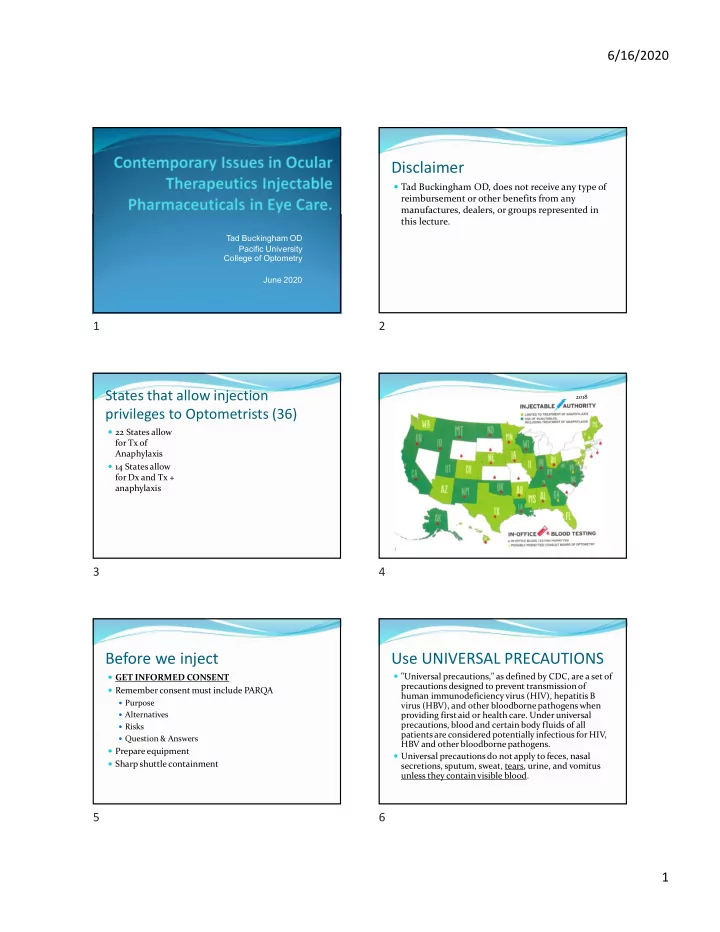
States that allow injection privileges to Optometrists (36)

 22 States allow for Tx of Anaphylaxis  14 States allow for Dx and Tx + anaphylaxis

January 2012 2018

Before we inject

 GET INFORMED CONSENT  Remember consent must include PARQA

 Purpose  Alternatives  Risks  Question & Answers

 Prepare equipment  Sharp shuttle containment

Use UNIVERSAL PRECAUTIONS

 "Universal precautions," as defined by CDC, are a set of precautions designed to prevent transmission of human immunodeficiency virus (HIV), hepatitis B virus (HBV), and other bloodborne pathogens when providing first aid or health care. Under universal precautions, blood and certain body fluids of all patients are considered potentially infectious for HIV, HBV and other bloodborne pathogens.  Universal precautions do not apply to feces, nasal secretions, sputum, sweat, tears, urine, and vomitus unless they contain visible blood.

1 2 3 4 5 6

slide-2
SLIDE 2

6/16/2020 2

UNIVERSAL PRECAUTIONS

 Universal precautions involve the use of protective barriers such as gloves, gowns, aprons, masks, or protective eyewear, which can reduce the risk of exposure of the health care worker's skin or mucous membranes to potentially infective materials. In addition, under universal precautions, it is recommended that all health care workers take precautions to prevent injuries caused by needles, scalpels, and other sharp instruments or devices.

Sharp shuttle examples Injection Applications

 Emergency Use

 Management of the acute allergic reaction Anaphylaxis  Rapid IOP reduction

 Diagnostic Use

 Fluorescein Angiography

 Therapeutic Use

 Local infiltration anesthesia

 Lesion (ie skin tag, chalazion) removal or structural repair via incision,

excision, or electrocautery  Steroid Deposition

 Chalazion, Scleritis, Iridocyclitis, and Recalcitrant Uveitis

 Antibiotic Deposition

 Non-Compliant patients, poor topical penetration, Increase focal

concentration

Local Block/Anesthesia Skin tag removal

7 8 9 10 11 12

slide-3
SLIDE 3

6/16/2020 3

Shave Biopsy Punch Biopsy

Conservative Chalazion treatment Warm compresses q.i.d. with gentle massage may resolve approx. 40% of small chalazions after several weeks. Oral antibiotic medications – Doxycycline 100mg qd x 2wks then 50mg qd x 2wks Z-pak (Azithromycin) 500mg PO q1d and 250mg qd x 4 days (total of 6 – 250mg tabs) Intralesional Steroid Injections Kenalog-40 (40mg/ml) with a 27-30 gauge needle in a 1cc syringe. Inject 0.1 – 0.2 cc into the lesion. Injecting the anterior lesion (skin side) does not require anesthesia. Use 1 gtt. topical anesthetic to numb up the injection site an reduce the blink reflex when injecting a posterior pointing (conjunctival) chalazion Resolution occurs in 1-2 weeks after one injection but 25% of chalazions may require a second injection in one month. Resolution of the chalazion is about 80% with just steroid injections and 90% when combined with conservative treatment  Chalazion Removal (Anterior pointing)  STEPS  Lid anesthesia (2% Lidocaine with epinephrine) 2 to 3cc with a 27-gauge needle. Massage. 

  • 1gtt. Ocular anesthesia to stop blink reflex

 Place Chalazion Forcepts(clamp) firmly.  Incise Chalazion using a sharp blade (#11) making a linear incision. Incise until lipomatous material presents.  Perform curettage.  Excise chalazion pseudocyst.  Remove clamp and perform hemorrhage control (direct pressure or use present clotting factors).  Prophylactic antibiotic ung. q.i.d. x 4 days. RTC in 1-2 weeks  Chalazion Removal (Posterior pointing)  STEPS  Lid anesthesia (2% Lidocaine with epinephrine) 2 to 3cc with a 27-gauge needle. Massage. 

  • 1gtt. Ocular anesthesia to stop blink reflex

 Place Chalazion Forcepts(clamp) firmly. Evert the lid.  Incise Chalazion using a sharp blade (#11) making a cruciate incision. Incise until lipomatous material presents.  Perform curettage.  Excise chalazion pseudocyst /capsule and truncate 4 corners of cruciate incision.  Remove clamp and perform hemorrhage control (direct pressure or use present clotting factors).  Prophylactic antibiotic ung. q.i.d. x 4 days. RTC in 1-2 weeks

Intralesional Injections Chalazion Incision and Curettage

13 14 15 16 17 18

slide-4
SLIDE 4

6/16/2020 4

Tad Buckingham OD, EMT-P

April 2019

Epinephrine 1:1000 (emergency)

 Epinephrine 1:1000 for Anaphylaxis  Anaphylaxis is an acute systemic (multi-system) and severe Type I Hypersensitivity allergic reaction in humans and other mammals. Minute amounts of allergens may cause a life-threatening anaphylactic

  • reaction. Anaphylaxis may occur after ingestion, skin

contact, injection of an allergen or, in rare cases, inhalation.

Anaphylaxis Diagnosis

Anaphylaxis Treatment

 Confirm the symptoms of Anaphylaxis

 Difficulty breathing  Feeling of airway constriction  Dizziness  Rash  Itching  Lowering blood pressure

 Call 911  Prepare Epinephrine 1:1000 (from MD vial or Ampule)

 Draw up 0.3 – 0.5ml in a 1 ml syringe (larger than 66 lbs)  Draw up 0.15ml in a 1 ml syringe (33 to 66 lbs)

 Administer IM or SQ  Repeat dose in 3-5 minutes if symptoms are not resolving

Anaphylaxis Treatment cont

OR  Call 911  Prepare Epinephrine 1:1000 by grabbing your weight appropriate Epi Auto-injector (Epi Pen)  Follow the directions of the Auto pen to deliver the epi (single dose) injection IM.  Prepare to give a second injection in 3-5 minutes if symptoms are not resolving.  NOTE: Pt. should be evaluated at an ED via ambulance immediately after the episode.

19 20 21 22 23 24

slide-5
SLIDE 5

6/16/2020 5

Anaphylaxis Treatment cont

 Antihistamine

 Diphenhydramine (Benadryl)

 Dose 50 mg IM, IV

Ondansetron (Zofran)

 Nausea/Vomiting Prevention  May be used during angle closure Glaucoma, if the patient is nauseous, and to prevent IOP spikes that occur during vomiting.  4mg IV/IM

 Can be repeated once if N/V is not controlled after 10 min.

 Contraindications

 Hypersensitivity to drug/class/componants  Hx of long QT syndrome  Caution with hepatic impairment  Caution with recent abdominal surgery

Acetazolamide (Emergency) IOP Reduction

 Most start with topical or oral medication. If IOP is still dangerously high add IV medications.  Acetazolamide 500mg/5ml IV

 Fast acting with peak onset of 15 minutes  Therapeutic duration of about 5 hours

 Contraindications

 Sulfa allergies (relative)  Pregnancy  Severe pulmonary obstruction  Severe Liver or Kidney disease  Low blood levels of K, Na or high blood levels of Cl

Fluorescein for Angiography

(Diagnostics)

 Fluroscein dye is injected into vein, from an established IV, to evaluate the retinal vasculature and it’s surrounding structures by using angiography.

 500 mg in 5 ml 10% sol. Or 2-3 ml 25% sol.

Anesthetics (Therapeutic)

 Lidocaine (Xylocaine)

 0.5, 1, 1.5, 2, 4% concentrations  1 minute onset with a maximum dose of 300mg  Duration is 30 to 60 minutes

 Lidocaine w/ Epinephrine

 1:100,ooo: 2% (20mg/ml) best ophthalmic concentration  1-5 minute onset with a maximum dose of 500mg  Duration of 2 to 6 hours

Corticosteroids (Therapeutics)

 Triamcinolone acetomide (Kenalog 40)

 Kenalog 40 works well, for intralesional injections,

because of the drug concentration (40mg/ml)

 Intralesional dose (ie. New onset Chalazions)

 0.5-30mg

 Methylprednisolone acetate (Depo-Medrol)

 Depo-Medrol 80mg/ml works well, for subconjunctival

injections, because of it’s concentration

 Subconjunctival dose (ie 2nd rnd Tx for recalcitrant

Uveitus and Iridocyclitis)

 40-80mg ( inject the dose in 2 to 4 subconjunctival sites)

25 26 27 28 29 30

slide-6
SLIDE 6

6/16/2020 6

Antibiotics (Therapeutic)

 Use Broad Spectrum Antibiotics unless cultures can be

  • identified. Subconjunctival antibiotic injections are

used in conjunction with different topical antibiotics  Broad Spectrum Antibiotic

 Cefazolin with Tobramycin/Gentamicin

 Subconjunctival dose 20mg in 0.5ml to 100mg in 0.5ml  Inject the dose close to the ulcer being treated

Injections supplies (what’s in the bag)

 Syringe

 1cc  5cc

 Tourniquet  Needles

 25ga  27ga  Winged Infusion Set  Bandaids  Cotton Balls  Tape  Gauze  0.9% Sodium Chloride Inj. USP  Alcohol Preps

Needle Taxonomy

 Needle Gauge

 External diameter  Larger the number the smaller the

diameter  Needle length

 Measured in inches

 Color

 Disposable needle color

Types of Injections

 Away from the Eye

 Deep Subcutaneous (SQ)  Intramuscular (IM)  Intravenous (IV)

 In or around the Eye

 Dermal  Subconjunctival (SC)  Intralesional  Peribulbar

Injections Away From the Eye

31 32 33 34 35 36

slide-7
SLIDE 7

6/16/2020 7

Deep Subcutaneous (SQ)

 Injection of up to 2 ml of solution deep under the skin but not into the muscular tissue.  Application

 Injection of 1:1000 epinephrine for acute treatment of

anaphylaxis  Equipment

 Alcohol wipes

Syringe (1ml-3ml) 23-25 gauge needle in adults/older children; 25 or 27g in infants 5/8 to 3/4 inch length sufficient; 1 inch optional length Substance to be injected Sterile gauze sponge Dressing (bandage or cotton and tape)

Deep Subcutaneous (SQ) cont.

 Location

 The fatty tissue over the biceps/triceps muscle near the deltoid muscle.

 Technique

 The injection site is rubbed vigorously with a swab and disinfectant to cleanse the area and increase the blood supply.  Make sure you pinch up on the SQ tissue to prevent injection into the muscle.  Insert needle, quickly, at a 45° angle into the fatty tissue over the triceps muscle.  The syringe should be aspirated a little by pulling back on the piston. If blood is present you are in a vein and the needle should be re-injected, and the piston withdrawn slightly once more.  The syringe piston is pushed down steadily and slowly.  A sterile cotton swab should be pressed over the injection site as the needle is quickly withdrawn, and the swab is taped to the skin for a few minutes, if required.

Deep Subcutaneous (SQ) Intramuscular (IM)

 Injection of up to 2 ml of solution into each muscular tissue site.  Application

 Injection of Epinephrine 1:1000 for acute treatment of

anaphylaxis

 Injection of Diphenhydramine for acute treatment of

anaphylaxis

 Injection of Ondansetron (Zofran) for N/V management

during acute angle closure Glaucoma  Equipment

 Same as Subcutaneous injections

Intramuscular (IM) cont.

 Location

 The muscular tissue of the deltoid/thigh muscle.

 Technique

 The injection site is rubbed vigorously with a swab and disinfectant to cleanse the area and increase the blood supply.  Insert needle at a 90° angle to the skin with a quick thrust.  The syringe should be aspirated a little by pulling back on the piston. If blood is present you are in a vein and the needle should be re-injected, and the piston withdrawn slightly once more.  The syringe piston is pushed down steadily and slowly.  A sterile cotton swab should be pressed over the injection site as the needle is quickly withdrawn, and the swab is taped to the skin for a few minutes, if required

Intramuscular (IM)

37 38 39 40 41 42

slide-8
SLIDE 8

6/16/2020 8

Intravenous (IV)

 Into a Vein  Applications

 Injecting Fluorescein dye into a vein for Fluorescein Angiography  Emergent lowering of high IOP with angle closure glaucoma

 Equipment

 Alcohol wipes

Syringe (1ml-5ml) 19-21 gauge winged infusion (“Butterfly”) set 3/4 inch needle length sufficient; 1 inch optional length Substance to be injected Tape Sterile gauze sponge Dressing (bandage or cotton and tape) Assistant to aid setup

Winged (“Butterfly”) IV set up

Venous IV Sites  Finding a suitable vein  Arms are a great place

 Place a tourniquet around the

upper arm

 Look and Feel for evidence of

the vein

 Encourage the pt to keep their

hand below their heart and also repeatedly make a fist with their hand and then relax it.

Is the tourniquet too tight????

 The tourniquet is on to tight if you cannot feel a pulse below the site of the tourniquet.  If you have the tourniquet on the upper arm check for the radial pulse

Intravenous (IV)

 Technique

 Take body substance isolation precautions (GLOVES)  Prepare needed equipment

 Set up Butterfly IV tubing  Syringe  Alcohol preps  Cotton balls and tape  Sharps container  Tourniquet

 Apply tourniquet around upper arm  Locate an appropriate IV site

Intravenous (IV)

 Technique (cont.)

 Clean site with alcohol prep.  Grab the wings of the Butterfly with the thumb and

forefinger in a pinching fashion

 Insert the needle into the vein “like an airplane landing on a

runway” with the bevel up.

 When you see a “flash back” (blood flowing from the vein,

through the needle, and into the IV tubing) while inserting the needle stop . You are in the vein.

 Remove the tourniquet  Tape the butterfly and tubing.  Flush the IV with normal saline.  Prepare for the IV injection.

43 44 45 46 47 48

slide-9
SLIDE 9

6/16/2020 9

Intravenous (IV)

Injections in and around the Eye

Dermal

 Injection of up to 2 ml of solution under the skin but not into the muscular tissue.  Applications

 Local infiltration anesthesia

 Lesion removal or structural repair via incision, excision, or

electrocautery

Dermal cont.

 Equipment

 Alcohol wipes

Syringe (1ml-3ml) 25-30 gauge needle 5/8 to 3/4 inch length sufficient; 1 inch optional length Substance to be injected Sterile gauze sponge Dressing (bandage or cotton and tape)  Location

 Upper and lower eye lids. Subcutaneous skin located in

the adnexa

Upper and Lower Eye lid injections

Eyelids Pearls When injecting anesethia in the eyelids : 1) Inject above the location on the upper eyelid 2) Inject below the location on the lower eyelid 3) Inject near the closest nerve This ensures that you achieve nerve block by following the anatomy of the nerves in the lower and upper eyelids

Dermal

49 50 51 52 53 54

slide-10
SLIDE 10

6/16/2020 10

Subconjunctival Injections

The injection of medications under the conjunctiva into the subconjunctival space  Applications

 Delivery of corticosteroids and antibiotics

 Equipment

 Topical anesthetic  Alpha Agonist (subconjunctival hemorrhage control)  Syringe (1ml TB)  27-30 gauge needle  Substance to be injected  1gtt of a topical broad spectrum antibiotic

Subconjunctival Injections cont.

 Location

 Subconjunctival space

 Inferior conjunctiva is the easiest to access and has the fewest patient

complaints

 Technique

 1-2gtt of topical anesthetic on the eye  1-2gtt of Alpha Agonist on the eye 1-2 minutes post topical

  • anesthetic. When the globe appears white you are ready.

 Perforate the conjunctiva with the syringe needle. You should

always be able to see the tip of the needle as it is inserted to the most distal medication depot site.

 Move the needle slightly. There should be no corresponding

globe movement.

 Inject 0.2-0.5ml of medication as you withdraw the needle

from the subconjunctival space.

 Add 1 gtt of topical broad spectrum antibiotic if applicable

Subconjunctival Injections

Intralesional Injections

 The direct delivery of medication percutaneously into skin lesions with minimal systemic effects.  Application

 Intralesional injection of corticosteroids

 Equipment

 Alcohol wipes

Syringe (1ml TB) 25-27 gauge needle Substance to be injected (Kenalog-40 ideal for chalazions) Chalazion forceps Sterile gauze sponge Dressing (bandage or cotton and tape)

Intralesional Injections cont

 Location

 Injection directly into the lesion

 0.1-0.2 ml dose is needed with Kenalog-40

 Technique

 Chalazions may be injected from the skin side or the

conjunctiva side –Use Chalazion forceps as a “Back stop” to the injection

 Skin side

 Sterilize the site  No anesthesia needed  Insert the needle into the center of the chalazion. Draw back on the

plunger of the syringe to confirm you are not in a blood vessel

 Inject the dose directly into the center of the lesion

Intralesional Injections cont

  • Technique cont.

 Conjunctiva side

 Anesthesia (1-2 gtt) is given to suppress the blink reflex and

numb the injection site

 Insert the needle into the center of the chalazion. Draw back

  • n the plunger of the syringe to confirm you are not in a blood

vessel

 Inject the dose directly into the center of the lesion

55 56 57 58 59 60

slide-11
SLIDE 11

6/16/2020 11

Intralesional Injections

Peribulbar Injections

Developed in 1986 as an alternative to retrobulbar injections The direct injection of anesthesia in the peribulbarspace  Application

 General ocular anesthesia for procedures such as RK,

cataract surgery, and vitreoretinal surgeries .  Equipment

 Sterilization solution for injection sites  27 gauge needle and 1-3 ml syringe  7/8 inch 23 gauge retrobulbarstyle needle and 6 ml syringe

Peribulbar Injections cont

 Location

 1st injection is in the lower eyelid fornix just above the inferior

  • rbital rim, approximately one finger breadth medial to the

lateral canthus.

 *If needed: a 2nd injection can be given in the inferonasal

  • rbit. The anesthetic is deposited below the globe inferior to

the lower punctum. The direction is not towards the globe, but slightly superior and posterior so as to avoid the globe.

 * If needed: a 3rd third injection can be given through the

upper lid below the superior orbital notch at the level of the lid crease - following basically the same technique as the 1st injection.

Peribulbar Injections cont

 Technique

 Inject a 0.5 ml wheal of lidocaine is raised at the 1st injection site.  An additional 0.5ml is then injected into the orbicularis oculi muscle

under the skin wheal, and approximately 1.0ml is deposited just anterior to the equator.

 A folded 4 X 4 is placed over the injection sites for approximately thirty

seconds to one minute to decrease potential ecchymosis.

 Using s standard 7/8 inch 23 gauge retrobulbar needle is inserted

transcutaneously just above the inferior orbital rim 1.5cm medial to the lateral canthus. The needle is then advanced along the inferior orbit to the equator of the globe. After aspirating to ensure that an intravascular injection will not be given, an additional 2.0ml of solution is deposited just anterior to the equator of the globe. The barrel of the syringe is then angled over the zygomatic arch and the needle is advanced in a slightly superior direction to approximately 7/8". After aspirating, 4.0ml to 6.0m1 of the solution is deposited just posterior to the equator.

Peribulbar Injections cont

 Technique cont.

 Note: After this injection the globe moves slightly up-

ward and minimally proptosis.

 At 8 minutes post injection test for akinesia and

anesthesia.

 If the akinesia and anesthesia are not 100% repeat steps

at the 2nd site. Evaluate after 8 minutes. If complete block is not noticed (RARE) repeat steps at the 3rd site.

Peribulbar Injections

Location of Injections

Always aspirate before the injections are given

1st 2nd 3rd

61 62 63 64 65 66

slide-12
SLIDE 12

6/16/2020 12

Peribulbar Injections

 Advantages of peribulbar Injections

 Easily learned technique  Less pain upon injection  No temporary vision grey or black-out as seen with

retrobulbar injections.

 Less posterior pressure problems compared with

retrobulbar injections

Recommended non-inclusive supply list to start using injectables in eye care:

Supplies

 Sharps container  Latex-free exam gloves  25 ga. 5/8 inch needle  27 ga. 5/8 inch needle  30 ga. ½ inch needle  19-21 ga. ¾ inch winged “butterfly” IV set  3cc disposable syringes  #11 disposable scalpels  Sterile cotton tipped applicators  Alcohol preps.  2x2 gauze  Tape  Cotton balls  Eye pads

Recommended non-inclusive supply list

to start using injectables in eye care:

Equipment Pharmaceuticals

 Chalazion Forcepts (clamps)  Chalazion curettes  Jeweler’s forcepts  Mouth-tooth forcepts  Small Wescott scissors  Disposable hand-held heat cautery unit  Head Loupe  Sterilization equipment/solutions  Lidocaine 2% with Epinepherine 1:100,000  Kenalog 40  Epinepherine 1:1000 MD vial/ampule or auto-injector

Any Questions?

67 68 69 70