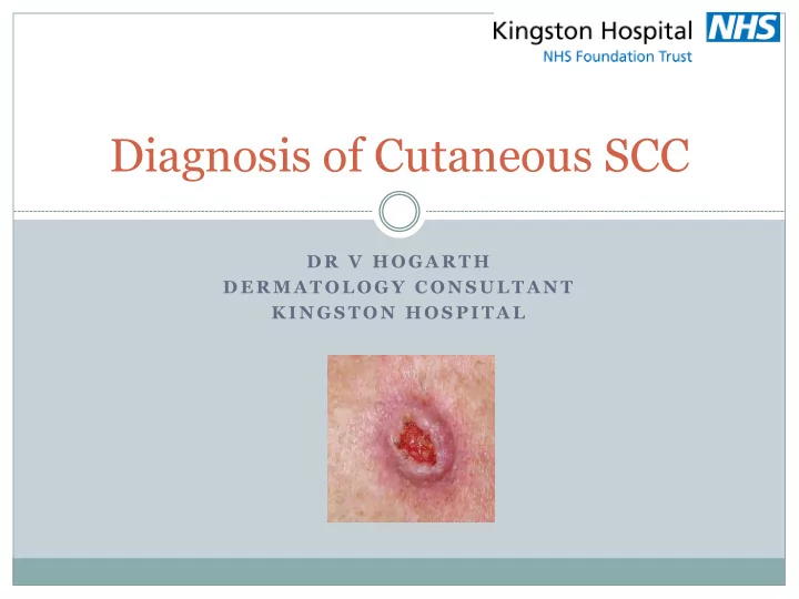
SLIDE 1 D R V H O G A R T H D E R M A T O L O G Y C O N S U L T A N T K I N G S T O N H O S P I T A L
Diagnosis of Cutaneous SCC
SLIDE 2 INTRODUCTION
Aim : Increased confidence screening for SCC Encourage follow up care in the community
especially for low risk tumours
1.
Diagnosis
3.
Follow up/Screening
SLIDE 3 Importance of SCC’s
1.
NMSC is the most common group of cancers
- 2. 23% of these are SCC’s
3.
Continuing increase in prevalence
- 4. Cancer of cells producing keratin
5.
May metastasise so treat early
SLIDE 4
SLIDE 5
Risk factors
SLIDE 6
SLIDE 7
SLIDE 8
SLIDE 9
SLIDE 10
Immunosuppression
HIV Immunosuppressive drugs Blood malignancies Organ transplant Receiving radiation These are more aggressive and greater risk of
metastasis
Previous cutaneous injury – longstanding
ulcer/thermal injury
SLIDE 11
History
Varies Enlarging over weeks to months Rapidly growing with pain and tenderness Pain is important because it can indicate perineural
invasion
SLIDE 12
Examination
Size Location ?recurrent ?connected to underlying structures Borders – well/poorly defined ?evidence of previous surgery Lymph nodes Full skin examination
SLIDE 13 Location
Within a background of
sun-damaged skin
Sun exposed sites 1.
Bald scalp
- 2. Face
- 3. Neck
- 4. Extensor forearms
- 5. Dorsal hands
- 6. Shin
SLIDE 14
A few mm Flesh coloured Several cm Erythematous
Size and colour variable
SLIDE 15
Initially thickening of skin and becomes an indurated plaque
SLIDE 16 Papulonodular
Gradually becomes fixed and nodular
SLIDE 17
Papulonodular
SLIDE 18 Keratin/’Crater’
Margin firm and more raised than a basal cell carcinoma, often everted and irregular
SLIDE 19
Plaque like
SLIDE 21
Warty/keratin horn
SLIDE 22
Poorly differentiated
SLIDE 23
SLIDE 24
SLIDE 25
Arising within ulcers
SLIDE 26
High risk features
Ears, lips, genitalia and other mucosal sites Greater than 2cm Tumour thickness greater than 4mm Moderate or poorly differentiation Perineural invasion Recurrent Arising form a scar or ulcer Lymphovascular invasion Immunosuppression
SLIDE 27
SLIDE 28
Thank you
SLIDE 29 Risk factors
UV radiation – SCC relate to chronic exposure (outdoor
Sun – lived abroad>holidays Sunbeds Fair skin, red hair, blue eyes Family history Immunosuppression (HIV, drugs, blood malignancies,
- rgan transplant, receiving radiation)
BCC/AK’s Smoking Previous cutaneous injury – longstanding ulcer/thermal
injury
SLIDE 30
Examination
A Less commonly plaque-like or warty Can be hyperkeratotic with thick crust Secondary changes include erosions and ulceration