Outline Kilovoltage unit definition Calibration and commissioning - - PDF document

outline
SMART_READER_LITE
LIVE PREVIEW

Outline Kilovoltage unit definition Calibration and commissioning - - PDF document

Outline Kilovoltage unit definition Calibration and commissioning of an PRACTICAL ASPECTS OF COMMISSIONING AND orthovoltage unit CALIBRATION OF CLINICAL ORTHOVOLTAGE UNITS Clinical applications Wamied Abdel-Rahman 1 , Li Heng


slide-1
SLIDE 1

McGill

Practical Medical Physics - AAPM 2009 1

PRACTICAL ASPECTS OF COMMISSIONING AND CALIBRATION OF CLINICAL ORTHOVOLTAGE UNITS

Wamied Abdel-Rahman1, Li Heng Liang2, Ismail Aldahlawi1, and Jan Seuntjens3

1) Montreal General Hospital, Montreal, Quebec, Canada 2) SMBD Jewish General Hospital, Montreal, Quebec, Canada 3) McGill University, Montreal, Quebec, Canada

McGill

Practical Medical Physics - AAPM 2009 2

  • Kilovoltage unit definition
  • Calibration and commissioning of an
  • rthovoltage unit
  • Clinical applications
  • Quality assurance

Outline

McGill

Practical Medical Physics - AAPM 2009 3

Low energy photon radiotherapy

(Based on F.M. Khan, The physics of radiation therapy, second Edition )

  • Grenz-ray therapy

– energy < 20 kVp

  • Contact therapy

– 40-50 kVp – SSD < 2 cm – ~ 2 mA

  • Superficial therapy

– 50-150 kVp – SSD = 15-20 cm – ~ 5-8 mA

  • Orthovoltage therapy (“Deep” therapy)

– 150-500 kVp – SSD ~ 50 cm – 10-20 mA

McGill

Practical Medical Physics - AAPM 2009 4

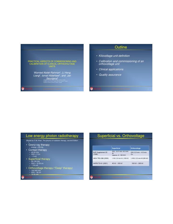
Superficial vs. Orthovoltage

Superficial Orthovoltage BJR Supplement 25 (1996) HVL 0.01 mm -8.0 mm Al (approx. 6 -150 KV) HVL 0.5 mm – 4.0 mm Cu IAEA TRS-398 (2000)

≤ HVL 3.0 mm Al (100 kV)

≥ HVL 2.0 mm Al (80 kV) AAPM TG-61 (2001) 40 kV - 100 kV 100 kV – 300 kV

slide-2
SLIDE 2

McGill

Practical Medical Physics - AAPM 2009 5

  • Skin cancer

– Melanoma – Basal cell carcinoma (BCC) – Squamous cell carcinoma (SCC)

  • Other skin lesions

– Keloid treatment

Treatment applications

McGill

Practical Medical Physics - AAPM 2009 6

  • Low energy photons

– Advantages

  • Sharp penumbra
  • Small lesions
  • Less complexity of machine and treatment
  • Easy setup

– Disadvantages

  • Penetrating beam
  • Higher dose to the bone
  • Electron beam advantages:
  • Sharp falloff of the PDD
  • Better cosmetic outcome
  • Bone “sparing”
  • Brachytherapy advantages:
  • Better outcomes for some selected sites

Kilovoltage clinical application

PDD (FS=10x10 cm 2)

10 20 30 40 50 60 70 80 90 100 1 2 3 4 5 6 7 8 9 10

Depth (cm) PD D (% )

6 MeV, SSD=100 cm 9 MeV, SSD=100 cm 12 MeV, SSD=100 cm 50 kVp, SSD=22 cm, cone = 3 cm diam, 0.25 mm Al 80 kVp, SSD= 40 cm, 2.45 mm Al 120 kVp, SSD= 40 cm, 3.75 mm Al 250 kVp, SSD= 40 cm, 2.02 mm Cu Co-60, SSD = 80 cm

McGill

Practical Medical Physics - AAPM 2009 7

Calibration and commissioning of an

  • rthovoltage unit

McGill

Practical Medical Physics - AAPM 2009 8

  • Gulmay Medical Limited, Chertsey, Surrey, UK

– Floor mounted tube stand and treatment table – Comet MXR321 metal Ceramic x-ray tube assembly – 9 treatment filters and 1 warm up filter – 6 square applicators (SSD = 50 cm) – 4 circular applicators (SSD = 20 cm)

  • X-ray beam specification

– X-ray tube output limits:

  • 20-220 kV, 0-20 mA, 400-3000 W
  • kVp at the JGH : 80, 120, 180, and 220 kVp
  • Tube

– Focal spot : ~7.5 mm – Target: Tungsten – Inherent filter: 0.8 ± 0.1 mm Be – Tube power max : 3000 W – Field coverage total: 40o – Anode angle: 30o – Weight: 11 Kg

Gulmay D3225 Orthovoltage Unit

slide-3
SLIDE 3

McGill

Practical Medical Physics - AAPM 2009 9

Square (cm x cm) SSD = 50 cm 4x4 6x6 8x8 10x1 15x1 5 20x2 Circular diameter (cm), SSD = 20 cm 3 4 5 10

Filters (9 + 1)

Gulmay D3225 Orthovoltage Unit McGill

Practical Medical Physics - AAPM 2009 10

Calibration

  • Protocol:

– The American Association of Medical Physicists Task Group 61 (AAPM TG-61)

  • Requirements

– Ionization chamber with an air kerma free in air calibration coefficient Nk traceable to national standards – NK can be calculated from the exposure calibration coefficient NX : NK = NX (W/e)air / (1-g)

McGill

Practical Medical Physics - AAPM 2009 11

Beam quality

  • Beam quality depends
  • n:

– Tube potential – Target material and angle – Window material and thickness – Monitor chamber material and thickness – Filtration material and thickness – Shape of collimator – Source-chamber distance

The physics of radiology, Johns & Cunningham

McGill

Practical Medical Physics - AAPM 2009 12

Beam quality specifier

  • Standards lab: HVL1 and kVp for

determination of Nk

  • Clinical unit: HVL1 for determination other

parameters in the TG-61 formalism

slide-4
SLIDE 4

McGill

Practical Medical Physics - AAPM 2009 13

Formalism: TG-61

  • In air method:

– Measurement performed in air – 40 kV ≤ Tube potential ≤ 300 kV

  • In phantom method:

– Measurement performed in Water – Size: At least 30×30×30 cm3 – 100 kV < Tube potential ≤ 300 kV

Ionization chamber

McGill

Practical Medical Physics - AAPM 2009 14

Dosimeters

  • Parallel-plate chambers:

– below 70 kV

  • Cylindrical chambers

– For 70 kV – 300 kV

  • Appropriate buildup should be used to eliminate

the effect of contaminating electrons

McGill

Practical Medical Physics - AAPM 2009 15

Determination of HVL

  • 1st HVL: thickness of a specified attenuator

that reduces the air-kerma rate in a narrow beam to ½ its original value.

– Measurement of the variation with the attenuator thickness of the air-kerma rate at a point in a “scatter free” and narrow beam. – Detectors with sufficient build-up should be used to eliminate the effect of contaminating electrons.

McGill

Practical Medical Physics - AAPM 2009 16

Determination of HVL: TG-61 recommendations

  • Diaphragm

– Beam diameter ≤ 4 cm – Thickness must attenuate primary beam to 0.1 %.

diaphragm 50 cm 50 cm Ionization chamber (detector) Monitor chamber Attenuator material

slide-5
SLIDE 5

McGill

Practical Medical Physics - AAPM 2009 17

Determination of HVL: TG-61 recommendations

  • Monitor chamber

– Used to correct for kerma rate variations – Should not perturb the narrow beam. – Should not be affected by the attenuator material

diaphragm 50 cm 50 cm Ionization chamber (detector) Monitor chamber Attenuator material

McGill

Practical Medical Physics - AAPM 2009 18

Determination of HVL: TG-61 recommendations

  • Attenuator

– High-purity material (99 %). – Thickness measured with an accuracy of 0.05 mm.

diaphragm 50 cm 50 cm Ionization chamber (detector) Monitor chamber Attenuator material

McGill

Practical Medical Physics - AAPM 2009 19

HVL measurement

McGill

Practical Medical Physics - AAPM 2009 20

HVL measurement

slide-6
SLIDE 6

McGill

Practical Medical Physics - AAPM 2009 21

Determination of Nk for a clinical beam (TG-61 recommendation)

  • Use of kVp and HVL

– Ideal: Obtain Nk for the same kVp and HVL beam that matches the user clinical beam – Practical: Obtain Nk for two beams with the same kVp but two HVLs and interpolate using the HVL for the clinical beam

McGill

Practical Medical Physics - AAPM 2009 22

Determination of Nk in the clinic (TG-61 recommendation)

  • ADCL’s provide

calibration coefficients for specific beam qualities that are grouped into:

– Lightly filtered (L series) – Medium filtered (M series) – Heavily filtered (H series)

  • Interpolation based on

HVL may only be performed within the same series

McGill

Practical Medical Physics - AAPM 2009 23

Calibration: Setup

  • Chamber: NE 2571 farmer type

cylindrical chamber

  • Method: in-air
  • Output specification point:

– 0 cm depth in water at the cone end – Inverse square factor is required for closed end cones to correct for chamber position

McGill

Practical Medical Physics - AAPM 2009 24

Percentage depth dose

  • Measurement medium:

water

  • Instrument: NACP01

parallel plate chamber

  • Window thickness = 90

mg/cm2

  • Electrode spacing = 2 mm
  • Effective point of measurement

= 1.9 mm

slide-7
SLIDE 7

McGill

Practical Medical Physics - AAPM 2009 25

PDD VS SSD PDD vs. SSD

McGill

Practical Medical Physics - AAPM 2009 26

Inline Crosslin e

Beam profiles: Inline and crossline

McGill

Practical Medical Physics - AAPM 2009 27

Back scatter factors

McGill

Practical Medical Physics - AAPM 2009 28

Dose to tissue

slide-8
SLIDE 8

McGill

Practical Medical Physics - AAPM 2009 29

Clinical application

McGill

Practical Medical Physics - AAPM 2009 30

SS D

1 mm SSD error

SSD (cm) SSD (cm) 20 20 50 50 ISL ISL (20.1/20) (20.1/20)2

2=1.010

=1.010 (50.1/50) (50.1/50)2

2=1.004

=1.004 Error(%) Error(%) 1.0% 1.0% 0.4% 0.4%

SSD vs potential errors

McGill

Practical Medical Physics - AAPM 2009 31

  • Entrance shielding: lead sheet, Cerrobend cutout
  • Exit shielding: lead, tungsten (eye shielding) (coated)

Custom Shielding

McGill

Practical Medical Physics - AAPM 2009 32

Cavity filling

slide-9
SLIDE 9

McGill

Practical Medical Physics - AAPM 2009 33

Quality assurance

McGill

Practical Medical Physics - AAPM 2009 34

Physics QA

Quality Assurance

McGill

Practical Medical Physics - AAPM 2009 35

Conclusions

  • The AAPM TG-61 is a protocol for reference dosimetry
  • f low energy photon beams (40 kV – 300 kV).
  • The effective point of measurement for parallel-plate

and cylindrical ionization chambers is the center of the sensitive air cavity.

  • If the dose close to or at the surface is of interest, the

in-air method should be used.

  • If the dose at depth is of interest, the in-phantom

method should be used.