SLIDE 1
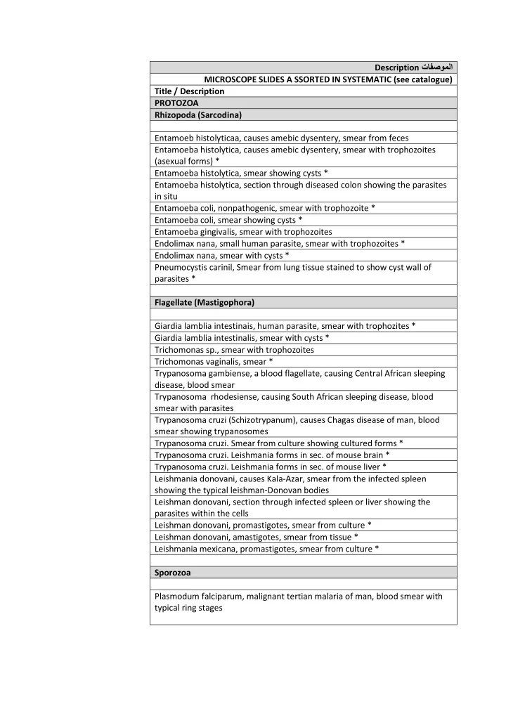
تافصوملا Description MICROSCOPE SLIDES A SSORTED IN SYSTEMATIC (see catalogue) Title / Description PROTOZOA Rhizopoda (Sarcodina) Entamoeb histolyticaa, causes amebic dysentery, smear from feces Entamoeba histolytica, causes amebic dysentery, smear with trophozoites (asexual forms) * Entamoeba histolytica, smear showing cysts * Entamoeba histolytica, section through diseased colon showing the parasites in situ Entamoeba coli, nonpathogenic, smear with trophozoite * Entamoeba coli, smear showing cysts * Entamoeba gingivalis, smear with trophozoites Endolimax nana, small human parasite, smear with trophozoites * Endolimax nana, smear with cysts * Pneumocystis carinil, Smear from lung tissue stained to show cyst wall of parasites * Flagellate (Mastigophora) Giardia lamblia intestinais, human parasite, smear with trophozites * Giardia lamblia intestinalis, smear with cysts * Trichomonas sp., smear with trophozoites Trichomonas vaginalis, smear * Trypanosoma gambiense, a blood flagellate, causing Central African sleeping disease, blood smear Trypanosoma rhodesiense, causing South African sleeping disease, blood smear with parasites Trypanosoma cruzi (Schizotrypanum), causes Chagas disease of man, blood smear showing trypanosomes Trypanosoma cruzi. Smear from culture showing cultured forms * Trypanosoma cruzi. Leishmania forms in sec. of mouse brain * Trypanosoma cruzi. Leishmania forms in sec. of mouse liver * Leishmania donovani, causes Kala-Azar, smear from the infected spleen showing the typical leishman-Donovan bodies Leishman donovani, section through infected spleen or liver showing the parasites within the cells Leishman donovani, promastigotes, smear from culture * Leishman donovani, amastigotes, smear from tissue * Leishmania mexicana, promastigotes, smear from culture * Sporozoa Plasmodum falciparum, malignant tertian malaria of man, blood smear with typical ring stages
SLIDE 2
تافصوملاDescription Plasmodum falciparum, blood smear with more gametocytes * Plasmodum vivax, benign tertian malaria of man, blood smear * Plasmodum vivax, thick diagnostic blood smear * Plasmodum sp. section through infected mosquito stomach with oocysts containing sporozoites * Plasmodium sp., section through the salivary gland of infected mosquito with sporozoites * Plasmodium sp., Exoerythrocytic stages in sec. of brain * Plasmodium sp., Exoerythrocytic stages in sec. of liver * Malaria melanemia in human spleen, sec. showing pigment granules in endothelium and Kuffer's cells Toxoplasma gondi, causing toxoplasmosis, tissue smear with parasites * Toxoplasma gondi, section of the brain showing cysts with parasites * Ciliate (Infusoria) Balantidum coli, human parasites, smear with trophozoites * Balantidum coli, smear with cysts * Balantidum coli, in sec. of human intestine * PLATYHELMINTHES-FLATWORMS Tubellaria-Turbellarians Fasc iola hepatica, t.s. through the body Fasc iola hepatica, t.s. through two different body regions Fasc iola hepatica, ova w.m. Fasc iola hepatica, miracidia (free living larvae) w.m. * Fasc iola hepatica, redia w.m. * Fasc iola hepatica, cercaria w.m. * Fasc iola hepatica, metacercaria w.m. * Fasc iola hepatica, redia and cercaria in sec. through infected snail liver Fasc iola hepatica in bile ducts of liver, t.s. Schistosoma mansoni, causing bilharziosis, adult male w.m. Schistosoma mansoni, adult female w.m. Schistosoma mansoni, adult male and female in copula, w.m. and carefully stained for general study Schistosoma mansoni, t.s. of adult male and female Schistosoma mansoni, miracaria w.m. * Schistosoma mansoni, cercaria with bifurcate tail w.m. * Schistosoma mansoni, section through infected snail liver showing cercaria Schistosoma mansoni, ova in section of liver or intestine * Schistosoma mansoni, ova in feaeces w.m. Schistosoma haematobium, ova from urine sediment w.m. Schistosoma japonicum, ova in faeces w.m. * Schistosoma japonicum, adult male w.m. *
SLIDE 3 Schistosoma japonicum, adult female w.m. * Schistosoma japonicum, miracidia w.m. * Description تافصوملا Schistosoma japonicum, cercariae w.m. * Cestodes-Tapeworms Taenia pisiformis (T. settata), tapeworm of dogs, immature proglottids w.m. Taenia saginata, tapeworm, proglottids w.m. * Taenia saginata, selected mature proglottids w.m. * Taenia saginata, t.s. of proglottids in different stages, the standard slide for general study Taenia saginata, ova in faeces w.m. Taenia solium, human tapeworm, proglottids t.s. Taenia solium, scolex w.m. * Taenia solium, ova in faeces w.m. Echinococcus granulosus, scolices from cyst, w.m. Echinococcus granulosus, ova in faeces of dog w.m. Echinococcus multilocularis, cyst with scolices t.s. NEMATHELMINTHES-ROUNDWORMS Ascaris lumbricoides, roundworm of man, t.s. of adult female in region of gonads Ascaris lumbricoides, t.s. of adult male in region of gonads Ascaris lumbricoides, t.s. of adult male and female in region of gonads Ascaris lumbricoides, isolated muscle cells w.s. Ascaris lumbricoides, larvae in sec. of pig lung Enterobius vermicularis, w.m. of adult male * Enterobius vermicularis, w.m. of adult female Enterobius vermicularis, ova from faeces w.m. Enterobius vermicularis, sec. through human appendix with parasites in situ Ancylostoma duodenale, hookworm of man, adult male w.m. * Ancylostoma duodenale, adult female w.m. * Ancylostoma duodenale, w.m. of adult male and female on one slide Ancylostoma duodenale, t.s. of male and female Ancylostoma duodenale, ova w.m. Ancylostoma duodenale,rhabditiform larvae w.m. * Ancylostoma duodenale, filariform larvae w.m. * Trichinella spiralis, section of infected muscle with encysted larvae Trichinella spiralis, w.m. of muscle with encysted larvae Trichinella spiralis, calcified larva in muscles, w.m. Trichinella spiralis, adult male from intestine w.m. * Trichuris trichiura, whip worm, w.m. of adult male or female * Trichuris trichiura, ova in faeces w.m. Trichuris trichiura, sec. of infected colon showing the parasitic worm in situ Microfilaria, smear from bird lung with parasites w.m.*
- I. Microscopic anatomy and histology
SLIDE 4
Head and mouth parts, whole mounts Description تافصوملا Musca domestica, house fly, head and mouth parts with sucking tube w.m. Anopheles, malaria mosquito, head and mouth parts of male w.m. Anopheles, head and mouth parts of females w.m. Culex pipiens, mosquito, head and moth parts of male w.m. Culex pipiens, head and mouth parts of female w.m. Culex pipiens, moth parts of female, dissected and w.m. * Diving beetle, head of larva w.m. Extraintestinal digestion * Simulium, head of larva w.m. show filtering mouth parts