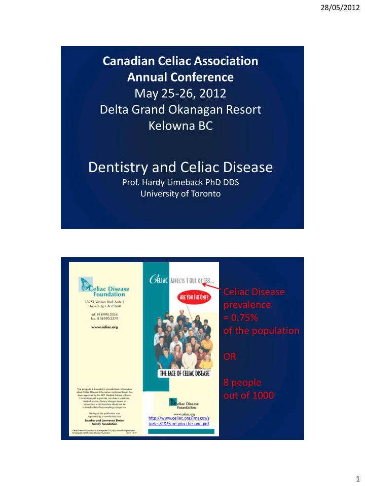
dentistry and celiac disease
play

Dentistry and Celiac Disease Prof. Hardy Limeback PhD DDS - PDF document

28/05/2012 Canadian Celiac Association Annual Conference May 25-26, 2012 Delta Grand Okanagan Resort Kelowna BC Dentistry and Celiac Disease Prof. Hardy Limeback PhD DDS University of Toronto Celiac Disease prevalence = 0.75% of the


  1. 28/05/2012 Canadian Celiac Association Annual Conference May 25-26, 2012 Delta Grand Okanagan Resort Kelowna BC Dentistry and Celiac Disease Prof. Hardy Limeback PhD DDS University of Toronto Celiac Disease prevalence = 0.75% of the population OR 8 people out of 1000 http://www.celiac.org/images/s tories/PDF/are-you-the-one.pdf 1

  2. 28/05/2012 Great Source of Information Only 3% to 5% of individuals with Celiac Disease are diagnosed Conditions that tend to mask or divert a diagnosis of celiac disease include dyspepsia, IBS, inflammatory bowel disease (IBD), tropical sprue, constipation, chronic fatigue, and various neurologic syndromes Source Celiac Disease Coeliac Disease, Celiac Sprue, Nontropical Sprue, Gluten-Sensitive Enteropathy Cara L Snyder, MS, CGC, Danielle O Young, MS, CGC, Peter HR Green, MD, and Annette K Taylor, MS, PhD, FACMG. 2

  3. 28/05/2012 J Clin Gastroenterol. 2010 Mar;44(3):191-4. www.celiacdiseasecenter.org Celiac Enamel Hypocalcification looks like dental fluorosis..... How can you tell the difference? Dr. Ted Malahias 3

  4. 28/05/2012 Kaukinen K, Collin P, Mäki M. 2007. Latent coeliac disease or coeliac disease beyond villous atrophy? Gut 56: 1339-1340 The Tooth Enamel 96% mineral Dentin 70% mineral Cellular Cementum ~ 65% mineral 4

  5. 28/05/2012 Mechanical and chemical destruction dental hard tissues Physical properties of Dental hard tissues -dentin has proteins, tubular spaces, small crystals and can flex -enamel is crystalline, has very large crystals and cracks easily… but is very hard 5

  6. 28/05/2012 Ultrastructure of Enamel Meckel et al., Arch. Oral Biol. 1965 • Enamel is the hardest, most highly mineralized biomaterial • >95% inorganic carbonated hydroxyapatite matrix • more resistant to fracture than geological apatite crystals (indicating how important biomineralization is) • Meshwork of interwoven crystal prisms that grow as ribbons/rods • Inter-rod space contains organic components Fincham et al., J.Struct. Biol. 1999 How enamel forms The developing tooth http://www.youtube.com/watch?v=5kRTtTYhtCU 6

  7. 28/05/2012 Enamel Formation: where things can go wrong • Lack of calcium (hypocalcemia, Vitamin D deficiency) • Enamel protein matrix malfunction (e.g. defective amelogenin) • Defective enzymes that remove enamel proteins (MMP20, kallikrein) • Injury to the developing tooth bud follicle Formation of Enamel Injury here = hypoplasia Injury here = hypocalcification 7

  8. 28/05/2012 Du et al., Science 2005 Fincham AG, et al (1994) J Struct Biol, 112, 103-109. Proteases in the Enamel Matrix Degradation of enamel proteins is required for crystal growth (mineralization) and is achieved by 2 enzymes • Enamelysin (matrix metalloproteinase-20; MMP-20) a Zinc-dependent metalloproteinase – MMP family (i.e. collagenase, gelatinase, stromelysin) – Expressed during late secretory and early maturation stage – Localized on chromosome 11q22 – KO mice show AI phenotype – Substrate specificity for amelogenins • Kallikrein-4 (KLK4, EMSP-1, prostase) – Serine proteinase family (i.e. trypsin, chymotrypsin) – Expressed during maturation stage – Gene located on chromosome 19q13 – Mutations linked with AI in humans 8

  9. 28/05/2012 Are CD patients deficient in zinc? YES • Tran CD, Katsikeros R, Manton N, Krebs NF, Hambidge KM, Butler RN, Davidson GP. Zinc homeostasis and gut function in children with celiac disease. Am J Clin Nutr. 2011 Oct;94(4):1026-32. NO • Botero-López JE, Araya M, Parada A, Méndez MA, Pizarro F, Espinosa N, Canales P, Alarcón T. Micronutrient deficiencies in patients with typical and atypical celiac disease. J Pediatr Gastroenterol Nutr. 2011 Sep;53(3):265-70. pH Oscillations During Enamel Maturation Smooth- Ruffle- ended ended Smith (1998) Crit Rev Oral Biol Med 9:128-161 Carbonic anhydrase is also essential for controlled mineralization 9

  10. 28/05/2012 What Do Celiac Teeth Look Like? Hypocalcification vs Hypoplasia Aguirre et al. (1997) Dental enamel defects in celiac patients. Oral Surg Oral Med Oral Path Oral Radiol Endod 87:646-650. 10

  11. 28/05/2012 Enamel defect classification in Celiacs (according to Aine et al, 1990) Hypocalcification Hypoplasia Hypoplasia Hypoplasia GRADE I 11

  12. 28/05/2012 GRADE II GRADE III 12

  13. 28/05/2012 GRADE IV Nikiforuk G, Fraser D. (1981) The etiology of enamel hypoplasia: a unifying concept. J Pediatr . Jun;98(6):888-93. 13

  14. 28/05/2012 http://www.smilemichigan.com/Portals/1/Jour nal%20Flash/October%202011/index.html 14

  15. 28/05/2012 Differential Diagnosis of Enamel Lesions Systemic and Local Factors D EVELOPMENTAL ENAMEL DEFECTS NOT RELATED TO CELIAC ENAMEL PROBLEMS Systemic -inborn errors of metabolism, -genetic problems galactosaemia, amelogenesis imperfecta, phenylketonuria, epidermolysis bullosa alkaptonuria, pseudohypoparathyroidism erythropoietic porphyria taurodontism primary hyperoxaluria heart disorders, -neonatal disturbances, unilateral facial hypoplasia hypertrophy premature birth -infectious diseases, hypocalcaemia Haemolytic anaemia -neurological disturbances, -endocrinopathies, Local -nutritional deficiencies, -trauma -nephropathies, -periapical osteitis (infected baby teeth) -enteropathies, -liver diseases Source: Pindborg JJ. Int Dent J. 1982 Jun;32(2):123-34. 15

  16. 28/05/2012 Mulberry molars Infection This is a congenital defect caused by syphilis. The occlusal surface of the molar has many small globules of enamel, not cusps. http://www.cbstraining.com/student/Dental/Oral Pathology/M.html http://oralpatho.blogspot.ca/2010/03/mulberr y-molar-and-mulberryand-custard.html Biliary Atresia An unusual disorder that creates the appearance of bluish green teeth. The congenital defect occurs during infancy whereby biliary atresia occurs, disrupting the draining of bile from the liver to the small intestine. Numerous developmental events can occur, depending on the extent of the condition. Not hereditary and not contagious. http://www.dental--health.com/bad_teeth_congenital.html 16

  17. 28/05/2012 http://www.dentistry.unc.edu/research/defects/pages/tdo.htm Individuals affected with TDO that have very thin enamel can benefit from covering the teeth with a bonded filled composite resin to help prevent exposure of pulp horns through wear. The enamel that is present is typically well mineralized and generally retains bonded materials adequately. Systemic insults are easy to detect: 1. Pairs of teeth are affected (both sides) 2. Teeth that erupt together, suffer together (e.g. Incisor-molar hypoplasia) It’s fun to play ‘Dr. House’ in the dental office. Doc: ‘You certainly had your hands full with your son being so sick at birth.’ Mom: ‘ How did you know? I forgot to mention that on the medical questionnaire.’ http://www.youtube.com/watch?v=5H40I25xR1w&feature=related 17

  18. 28/05/2012 Sequence of tooth formation and eruption http://www.youtube.com/watch?v=5H40I25xR1w&feature=related Linear Enamel Hypoplasia (LEH) (the primary incisors were affected during development right after birth) Return to normal enamel after birth Neonatal Hypoplasia Enamel development protected in utero LEH indicates severe hypocalcemia at birth 18

  19. 28/05/2012 Is breast feeding protective? Primary Central Lateral First Second teeth incisor incisor Canine molar molar calcification 14 wk I.U. 16 wk I.U. 17 wk I.U. 15.5 wk Initial 19 wk I.U. I.U. Birth Crown completed 1.5 mo 2.5 mo 9 mo 6 mo 11 mo Root completed 1.5 yr 2 yr 3.25 yr 2.5 yr 3 yr Ash, Major M.; Nelson, Stanley J. (2003). Wheeler's dental anatomy, physiology, and occlusion . Philadelphia: W.B. Saunders. pp. 32, 45, and 53. • Does CD cause enamel hypoplasia in primary teeth? • When is gluten usually introduced into the infant’s diet? • Does a wheat-based porridge cause enamel hypoplasia in the second primary molars in infants with CD? Lagerqvist C, Dahlbom I, Hansson T, et al. (2008) Antigliadin immunoglobulin A best in finding celiac disease in children younger than 18 months of age. J Pediatr Gastroenterol Nutr. 2008 Oct;47(4):428-35. Fig. 3 (Rashid et al) Possible scenario : Gluten was introduced into the diet at age 1.5 years (which caused the enamel defect) then a gluten-free diet was introduced a month later after it was discovered that the diet containing gluten was making the child sick. Liversidge HM. Crown formation times of human permanent anterior teeth. Arch Oral Biol. 2000 Sep;45(9):713-21. 19

  20. 28/05/2012 If this were a child with Celiac Disease, could he avoid dental defects in his anterior teeth by continuing to drink breast milk???? Differential Diagnosis of Enamel Lesions Ameogenesis Imperfecta 20

  21. 28/05/2012 Enamelin Translation Termination at Codon 53 Autosomal Dominant Local Hypoplastic AI Mårdh et al ., (2002) Hum Mol Gen 11:1069-74 APECED: Autoimmune Polyendocrinopathy- Candidiasis-Ectodermal Dystrophy Mutations in autoimmune regulator ( AIRE , 21q22.3) APECED causes multiple endocrine deficiencies, oral candidiasis and different forms of ectodermal dystrophy including enamel hypoplasia. Pavlic & Waltimo-Siren (2009) Arch Oral Biol 54:658-65 21

Download Presentation
Download Policy: The content available on the website is offered to you 'AS IS' for your personal information and use only. It cannot be commercialized, licensed, or distributed on other websites without prior consent from the author. To download a presentation, simply click this link. If you encounter any difficulties during the download process, it's possible that the publisher has removed the file from their server.

Recommend


More recommend