
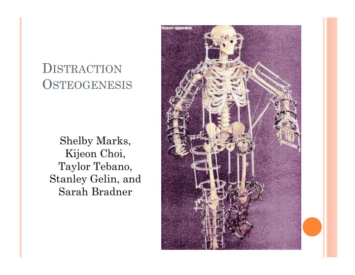
SLIDE 1
DISTRACTION OSTEOGENESIS
Shelby Marks, Kijeon Choi, Taylor Tebano, Stanley Gelin, and Sarah Bradner
D ISTRACTION O STEOGENESIS Shelby Marks, Kijeon Choi, Taylor - - PowerPoint PPT Presentation
D ISTRACTION O STEOGENESIS Shelby Marks, Kijeon Choi, Taylor Tebano, Stanley Gelin, and Sarah Bradner L EARNING O BJECTIVES To understand the advancement of bone elongation techniques that lead to modern day practice. Be able to
DISTRACTION OSTEOGENESIS
Shelby Marks, Kijeon Choi, Taylor Tebano, Stanley Gelin, and Sarah Bradner
LEARNING OBJECTIVES
To understand the advancement of bone elongation
techniques that lead to modern day practice.
Be able to describe what distraction osteogenesis is
and how it works.
Identify and compare the different distraction
reconstruction.
Understand the many causes/diseases for which
distraction osteogenesis is used.
Recognize the complications and risks associated
with the process and identify post-operative care.
DISTRACTION OSTEOGENESIS
Surgical reconstruction of skeletal
deformities and lengthening of the long bones of the body
Procedure that moves 2 bone segments apart
using an external saw and allows new bone in gap
Many FDA approved distraction devices
by Hippocrates (460-377 B.C.) when traction on long bones was performed by means of rubber straps
bone elongation techniques
extension using axial forces
had a high complication rate
Gavril Ilizarov
– Gavriil Abramovich Ilizarov was born in Caucasus (former USSR) in 1921 – No formal schooling till age 11 – Lone physician in Kurgan, Serbia – Developed Ilizarov method – An external fixator to regenerate bone under tension – modular ring – Died July 24 1992 in Kurgan, Russia due to heart failure
RUSSIAN ILIZAROV SCIENTIFIC CENTER FOR RESTORATIVE TRAUMATOLOGY AND ORTHOPAEDICS
managing bone infection
were first devised and introduced into clinical practice.
HOW DOES IT WORK?
Must be gradual
too quick: fibrous union instead of osseointegration too slow: early bone union Steps:
1)
Surgeon makes ostectomy (break) in abnormal bone and places distraction device. Allowed to start a few days of healing.
2)
Parent/ guardian turns a screw on distractor after surgery at home to stretch healing tissue (stretch~1mm/day)
3)
Stretch regenerated tissue forms into new bone
4)
After bone is formed at necessary length, device is removed through a 2nd surgery
CELLULAR LEVEL:
Aka callus distraction Inflammation: formation of
blood clot and ingrowth of angiogenic elements
Soft callus: replacement of
blood clot with granulation tissue and fibrocartilage
Hard Callus: granulation and
fibrocartilage is is replaced with woven bone
Remodeling: woven bone is
replaced with lamellar bone
RIGID external device (RED) Frames surgery Intramedullary Nail
LRS
Found to give reliable results, control bone
movements
easy removal Cranial halo (semicircular piece) encircles front
touch but do not penetrate bone.
Cranial halo is attached to vertical graphite
EXTERNAL DEVICE
Ex: Midface advancement
MIDFACE ADVANCEMENT
Make cut in bones of face at specific points Loosen midface from rest of skull Advance into correct position
New advantages:
needed with plates and screws
do not fight to push back
SURGERY”
Oldest and most common
method
Application of circular frame
Most common
1) Ilizarov frame 2) Taylor Spatial frame
Frames are circular external
fixators that surround limb
Consists of 4-6 rings made of
stainless steel or carbon fiber
Thin wires and half pins are fixed
to rings and pass through skin to bone Half pins: attached on one side of frame Wires: other side through bone and attached and held under tension Rods and struts: between rings for stability and adjustment
INTRAMEDULLARY NAIL
Internal telescopic nail with
lengthening mechanism
Lengthening achieved by
rotation
Rotation makes “click” noise Click allows higher
accuracy, patient knows how much they lengthened it
15 clicks~1 mm Fully implantable nail
allows for full weight bearing
4 inch gain Small scar
Improved method Stronger steel makes it weight bearing No longer “one size fits all” Can accommodate different sized limbs
FITBONE (FULLY INTEGRATED TELESCOPIC BONE)
Nail is distraction
device powered by internal engine
Engine activated by
hand remote
Activates distraction
by sending messages to receiver below skin
Similar function as car antenna Fitbone elongation is propelled by gear Inserted through “model entrance point” Tiny scar
Activated by polar movements through
small rotation of bone segment being lengthened
Amount of length is determined pre-op
and set at time of insertion
Allows 3’’ gain
SYSTEM)
Includes different clamps
that slide on rigid rail and connected with distraction units
Relies on callus
distraction
Stability by allowing
different bone screw position in clamp along length of bone
Rail length pick
depending on size of limb
Osteotite bone screws
with hydroxyapetite coating
Types of Congenital Deformities:
limb lengthening
facial deformities
amputation at the elbow
correction of airway obstruction in micrognathia
Fibular hemimelia
Hemihypertrophy
Olliers disease
Procedure:
a bone segment is surgically cute and a distraction device is used to slowly pull the two ends apart.
After the desired lengthening has been achieved, the bone consolidates until the lengthened gap has completely calcified.
Distraction phase and Consolidation Phase in distraction osteogenesis.
History: Discovered by Russian Orthopedic Gavril Ilizarov
Technique: external fixator
Today: minimally invasive techniques performed with internal distraction device
improve fitting of prostheses
Hemifacial Microsomia: a rare congenital disease
characterized by facial asymmetry
Cleft palate: separation of the roof of the mouth that
Severe mandibular hypoplasia (small lower jaw):
causes breathing problems includes conditions as:
Brachygnathia: abnormal shortness or recession of
the lower jaw
Micrognathia: an abnormally small, lower jaw and
chin.
Why?
Hemifacial Microsomia
Distraction Process
Bimaxillary distraction osteogenesis allows correction of mandibular asymmetry with simultaneous correction of the position of the maxilla
Cleft Palate
OBSTRUCTION
Micrognathia
Can use tracheostomy: May be life saving but is associated
with complications and developmental problems
Alternative: internal mandibular distraction osteogenesis
Treacher-Collins syndrome: a condition that affects the
development of bones and other tissues in the face
Pierre-Robin Sequence: a group of disorders occurring
together that includes a small, lower jaw, breathing problems, and a tongue that tends to ball up at the back of the mouth
Facial Injuries:
cause damage to the maxilla or mandible most common condition for which distraction osteogenesis is
performed
MAXILLOFACIAL DISTRACTION OSTEOGENESIS
Two Main Uses:
Craniofacial Defects
trauma/ impact
Requires making
bones longer
MAXILLOFACIAL DO PROCESS
Bonus Question: Why is it the hardest
to grow bone in the face considering the current methods?
MAXILLOFACIAL DO
Bone movements
must be carefully planned before a device is implanted
There are no devices
that can change trajectory mid-course
MAXILLOFACIAL DO PROCEDURES
Current Methods: External and linear
(one dimension)
Future device goals Curvilinear devices
(capable of moving bone in 3 dimensions)
Move bone
continuously, not in increments of 1mm
Causes less pain,
wouldn’t require patient compliance, might promote faster bone growth
Clavicle Lengthening Reconstruction of forearm
deformity
Foot deformities Bow legs
Clavicular lengthening by distraction osteogenesis for
congenital clavicular hypoplasia enables gradual correction of deformity
Has potential to improve shoulder pain, function, range
Deformity caused by osteochondromas corrected using an external
fixator for ulnar lengthening and radial deformity.
Why? Cosmetic and functional problems
Distraction osteogenesis used for the treatment
Condition in which there are one or more abnormally
short metatarsals, making the toe short as well
Blount’s Disease: In some cases, abnormal growth of the bone
causes the bowing to get worse instead of better over time
Caused by a growth disorder in the upper part of the tibial
bone
DISTRACTION
AFTER TUMOR RESECTION
After Bone Tumor
POST-TRAUMATIC INJURIES
Growth Plate
Fractures
Malunion Shortening and
deformity
Bone defects
INFECTION: OSTEOMYELITIS
Lansana’s Story:
13 year old boy from
Sierra Leone was bitten by a snake while chasing a soccer ball into a bush
Infection set in his tibia,
which led to a massive bone loss of his tibia and his ankle joint
Right leg was 6 inches
shorter then left, with a 45 degree internal rotation
COSMETIC USE
Short Stature Normal people that
are unhappy with their height
Dwarfism Achondroplasia
SHORT STATURE
Generally
Discouraged
Breaking perfectly
functioning limbs
Confining themselves to
wheelchairs or crutches for over a year
Voluntarily subjecting
themselves to pain and discomfort
Exposed to unnecessary
risk of infection, damaged nerves and blood vessels, and fat embolism
Expen$ive procedure Before procedure, they
must pass the body image assessment (US)
COSMETIC - CHINESE
National Geographic Video
DWARFISM
Achondroplasia:
Arms: Often times, the humerus
is much shorter than the radius and ulna
Forearm: More complex nerve
arrangement so few lengthenings are performed on that region
Legs: Femur and Tibia are
lengthened
Fibula is cut and
separated alongside the tibia
BEFORE & AFTER SURGERY
POST-SURGERY
Often prescribed painkillers and unable to work
while undergoing rehabilitation
Following initial surgery: Demanding Physical Therapy Purpose: Avoid stiffness and to stimulate the muscles, nerves,
and blood vessels to grow alongside the bone
WEIGHT BEARING
Depends on design of fixator and
bony contact
Volume of regenerate will affect
stability
Initially want strong support for
early weight bearing
Gradually remove wires and pins
so bone take on more load
MID FACE DISTRACTION: POST-SURGERY CARE
Hygiene – do not submerge device, soft
toothbrush, no mouthwashes
Diet – soft foods Medications – antibiotics, tylenol Activity – no sports, sitting and walking upright
good for healing/decrease swelling
THINGS TO LOOK
OUT FOR IN FACIAL
DISTRACTION
Persistant fever or
headache
Fall or blow to device Increased swelling
after initial post-op swelling
Axial deformity Pin tract infection Fixator instability Stress fractures in surrounding
PIN TRACT INFECTION
Disrupt blood supply Local inflammation Swelling
FIXATOR INSTABILITY
Excessive motion
in between bone segments
Premature
consolidation
NEW BONE PROPERTIES
Approx. 50% mechanical stiffness during axial,
torsional, and bending loading after fixator removal up to 6 weeks
Peak tensile forces increase with time after
completion
Torsional loads to failure decrease with time -
Why?
FORCES DURING DISTRACTION
Strain gauges Distraction load (resistance to
distraction) increases with every turn
Frictional forces – Where?
Good for: Humerus and femur Until recently – preference in U.S. Less awkward/more comfort Less bulk (Clothing
Protect skin and provide elevation Prevent cantilever bending Additional rings – more support Hinges for across joints
CIRCULAR FIXATOR - STABILITY
Smaller the diameter the better However not too close to skin, avoid
compression
Extra ring – no attachment to bone
but can be used to increase stability
VETERINARY CARE
Canine limb