cytometry
play

Cytometry Flow Cytometry Flow Cytometry is the technological - PowerPoint PPT Presentation

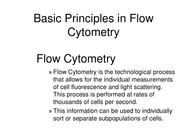
Basic Principles in Flow Cytometry Flow Cytometry Flow Cytometry is the technological process that allows for the individual measurements of cell fluorescence and light scattering. This process is performed at rates of thousands of cells


  1. Basic Principles in Flow Cytometry Flow Cytometry » Flow Cytometry is the technological process that allows for the individual measurements of cell fluorescence and light scattering. This process is performed at rates of thousands of cells per second. » This information can be used to individually sort or separate subpopulations of cells.

  2. History • Flow cytometry developed from microscopy. Thus Leeuwenhoek is often cited in any discussion regarding it’s history. • F.T. Gucker (1947)build the first apparatus for detecting bacteria in a LAMINAR SHEATH stream of air. • L. Kamentsky (IBM Labs), and M. Fulwyler (Los Alamos Nat. Lab.) experimented with fluidic switching and electrostatic cell sorters respectively. Both described cell sorters in 1965. • M. Fulwyler utilized Pulse Height Analyzers to accumulate distributions from a Coulter counter. This feature allowed him to apply statistical analysis to samples analyzed by flow.

  3. History • In 1972 L. Herzenberg (Stanford Univ.), developed a cell sorter that separated cells stained with fluorescent antibodies.The Herzenberg group coined the term Fluorescence Activated Cell Sorter (FACS).

  4. Fluorescence Activation Process (or Immunofluorescence) Antibodies recognize specific Antibodies are artificially molecules in the surface of conjugated to fluorochromes some cells FITC FITC Antibodies When the cells are analyzed by flow FITC cytometry the cells expressing the marker FITC for which the antibody is specific will manifest fluorescence. Cells who lack the marker will not manifest fluorescence But not others

  5. Cellular Parameters Measured by Flow Intrinsic Extrinsic • Reagents are required. • No reagents or probes required ( Structural ) – Structural – Cell size(Forward Light • DNA content Scatter) • DNA base ratios – Cytoplasmic grabularity(90 • RNA content degree Light Scatter) – Functional – Photsynthetic pigments • Surface and intracellular receptors. • DNA synthesis • DNA degradation (apoptosis) • Cytoplasmic Ca++ • Gene expression

  6. Flow Cytometry Applications • Immunofluorescence • Cell Cycle Kinetics • Cell Kinetics • Genetics • Molecular Biology • Animal Husbandry (and Human as well) • Microbiology • Biological Oceanography • Parasitology • Bioterrorism

  7. • Flow cytometry integrates electronics, fluidics, computer, optics, software, and laser technologies in a single platform.

  8. Sample Cells are presented Y Z to the laser using Sheath principles of hydrodynamic X focusing Flow chamber Y Z Laser optics X Laser Beam

  9. Laminar Fluidic Sheath Core Sheath PE FL Outer Sheath FITC FL 488nm Sct

  10. • Each cell generates a quanta of fluorescence Photomultiplier Tubes (PMT’s) PE FL FITC FL 488nm Sct Discriminating Filters Forward Light Dichroic Lenses Confocal Lens Scattering Detector

  11. Negative cells are also detected PE FL FITC FL 488nm Sct Forward Dichroic Lenses Light Scatter Confocal Lens

  12. Optical Bench Schematic FL3 Sensor FL4 FL2 620BP Sensor Sensor 675BP 575BP FL1 Sensor 525BP SS Sensor 645DL Fluorescence Pickup Lens 600DL 550DL Laser Beam 488BK 488DL Flow Cell FS Sensor

  13. From Fluorescence to Computer Display • Individual cell fluorescence quanta is picked up by the various detectors(PMT’s). • PMT’s convert light into electrical pulses. • These electrical signals are amplified and digitized using Analog to Digital Converters (ADC’s). • Each event is designated a channel number (based on the fluorescence intensity as originally detected by the PMT’s) on a 1 Parameter Histogram or 2 Parameter Histogram. • All events are individually correlated for all the parameters collected.

  14. Light Scattering , 2 Parameter Histogram Bigger Apoptotic Cells Bigger 90 degree Cells Light Scatter Dead Cells More Y Axis Granular X Axis Live Cells Forward Light Scatter (FLS)

  15. 1 Parameter Histogram Positive Negative Brighter Count Dimmer 6 4 1 1 2 3 4 6 7 150 160 170 .. 190 Channel Number Fluorescence picked up from the FITC PMT

  16. 2 Parameter Histogram Single Double Positive Positive PI Population Population PE FL Negative Population Single Positive FITC FL FITC Population

  17. Gating and Statistics • Data generated in flow cytometry is displayed using Multiparamater Acquisition and Display software platforms . • Histograms corresponding to each of the parameters of interest can be analyzed using statistical tools to calculate percentage of cells manifesting specific fluorescence, and fluorescence intensity. • This information can be used to look at fluorescence expression within subpopulations of cells in a sample (gating).

  18. Flow Cytometry Data Smaller Region, Live cells mostly Larger Region includes all cells

  19. Running Samples • Prepare samples. • One sample should be completely negative. This sample should be analyzed first. This sample is used for adjusting the PMT’s amplification voltage. • Adjust the PMT Voltage until you can see a population peak in the first decade of your 1 parameter and or your two parameter plot.These samples are used for adjusting Spectral Overlap . • Once the instrument settings are optimized, run samples and collect data.

  20. Flow Cytometry and sorting

Download Presentation
Download Policy: The content available on the website is offered to you 'AS IS' for your personal information and use only. It cannot be commercialized, licensed, or distributed on other websites without prior consent from the author. To download a presentation, simply click this link. If you encounter any difficulties during the download process, it's possible that the publisher has removed the file from their server.

Recommend


More recommend