current status of supplementary screening with breast
play

Current Status of Supplementary Screening With Breast Ultrasound - PDF document

Current Status of Supplementary Screening With Breast Ultrasound Stephen A. Feig, M.D., FACR Fong and Jean Tsai Professor of Womens Imaging Department of Radiologic Sciences University of California, Irvine School of Medicine Swedish


  1. Current Status of Supplementary Screening With Breast Ultrasound Stephen A. Feig, M.D., FACR Fong and Jean Tsai Professor of Women’s Imaging Department of Radiologic Sciences University of California, Irvine School of Medicine 
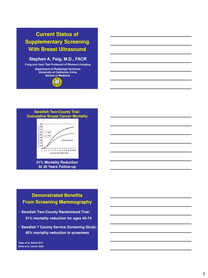
Swedish Two-County Trial: Cumulative Breast Cancer Mortality 31% Mortality Reduction At 30 Years Follow-up Demonstrated Benefits From Screening Mammography • Swedish Two-County Randomized Trial: 31% mortality reduction for ages 40-74 • Swedish 7 County Service Screening Study: 45% mortality reduction in screenees Tabar et al, Radiol 2011 Duffy et al, Cancer 2002 1

  2. Relative Likelihood of Interval Cancers Density Odds Ratio 95% CI < 10% 1.0 10-24% 2.1 (0.9 - 5.2) 25-49% 3.6 (1.5 - 8.7) 50-74% 5.6 (2.1 - 15.3) > 75% 17.8 (4.8 - 65.9) p < .001 Boyd et al New England J Med 2007;356:227-236 Can ultrasound find cancers missed by screening mammography? Breast Scanner developed in Australia, 1965 Courtesy, Jack Jellins Ph.D. 2

  3. Early Studies of Screening Ultrasound in 1980 ’ s • Inadequate detection of smaller cancers • Excessive false positive biopsies • Performance was time consuming • Expensive Improvements in Breast Ultrasound in 1990 ’ s • Better spatial resolution: 7.5 -10 MHz transducers • Better contrast resolution • Stavros criteria for interpretation Cancers Detected by Ultrasound Alone In Dense Breasts: 6 Screening Series, 1995 - 2003 • 150 cancers / 42,838 exams • 3.5 cancers / 1,000 exams • 90% in dense breasts • Mean tumor size of 0.9 – 1.1 cm • All Stage 0 or Stage I 3

  4. Increased Detection: Ultrasound and Mammography vs. Mammography Alone Study Increased Detection Kolb et al 1 42% Buchberger et al 2 37% Leconte et al 3 79% 1 Radiology 1998, 2002; 2 AJR, 1999; 3 AJR, 2003 False Positive Biopsies in Ultrasound Screening • 2.5 x – 4.0 x higher than mammography • Studies did not define biopsy criteria • Higher false positive rates likely with ultrasound screening in community practice Scientific Limitations of Screening Ultrasound Studies • Non-blinded ultrasound interpretation • Same radiologist read both modalities • No documentation of technical quality or interpretive expertise 4

  5. Multicenter Trial Protocol • Independent interpretation of ultrasound and mammography • Standardized ultrasound interpretive criteria • High resolution ultrasound equipment • Mammography and ultrasound technique monitored with quality control Multicenter Trial Protocol • Patients randomized to initial mammography or sonography • Ultrasound performed by radiologists • Radiologists: - received prior training in mammo and US interpretation - me t interpretive performance standards prior to participation High Risk Enrollment Requirements: At Least One of These Criteria • BRCA-1 or 2 mutation • Personal history of breast cancer • Biopsy proven - Lobular carcinoma in situ (LCIS) - Atypical ductal hyperplasia (ADH) - Atypical lobular hyperplasia (ALH) - Atypical papillary lesion • Prior radiation treatment of chest or axilla • Gail of Claus model risk of  25% 5

  6. Cancer Detection Rates at First Screening Round, ACRIN 6666 Trial: Hand-held Ultrasound Screening of High Risk Women • Mammography alone 7.6 / 1,000 • Mammography + US 11.8 / 1,000 • Supplementary yield for ultrasound 4.2 / 1,000 or 55.3 % increase Berg et.al. JAMA 2008 Biopsy Positive Predictive Value at First Screening Round, ACRIN 6666 Trial: Hand-held Ultrasound Screening of High Risk Women • Mammography with 22.6 % Ultrasound correlation • Ultrasound alone 8.9 % • Mammography or Ultrasound 11.2 % Berg et.al. JAMA 2008 Results at Second and Third Screening Rounds: ACRIN 6666 Trial • Supplementary yield of ultrasound = 3.7 cancers / 1,000 screens • Biopsy PPV: Mammography alone = 38% Mammo + ultrasound = 16% Berg et all, JA MA 2012; 307: 1394 - 1404 6

  7. Limitations of Screening with Hand-held Ultrasound • Exam time of 19 minutes (ACRIN Trial) • Technique / Interpretation are linked and operator-dependent • Need to document technologists’ skill for screening Significance of Screening Ultrasound Performance Time • Might lose money at screening mammography rates • Low reimbursement might encourage excessively fast screening times • Automated scanners might be the solution Follow-Up of Sonographic vs Mammographic Probably Benign Lesions • Sonographic follow-up is much more time consuming and operator dependant 7

  8. Methods to Facilitate Follow-Up of Probably Benign Ultrasound Lesions • Annual instead of 6 month follow-up • Development of a high resolution, automated whole breast ultrasound scanner ™ an Automated Breast Ultrasound System View Station Scan Station 8

  9. Advantages of Coronal View • New for breast ultrasound • See slices of entire breast from skin to chest wall • Tissue thickness reduced so better visualization 26 Invasive Ductal Carcinoma 9

  10. Benign Fibroadenoma Advantages of Automated Whole Breast Scanners • Rapid acquisition time of 10 minutes • Does not require physician performance • Allows batch reading • Can be integrated efficiently into breast center workflow Interpretive Aspects of Automated Breast Ultrasound (ABUS) • Suspicious findings may need hand-held confirmation and evaluation • Hand-held transducer required for ultrasound-guided biopsy • Some ABUS units have attached hand-held transducers 10

  11. Automated Scanner with Handheld Capability Increased Cancer Detection by Adding ABUS to DM For Screening Dense Breasts All Cancers 31% 19 / 62 DCIS 6% 2 / 31 Invasive Cancers 55% 28 / 51 Stage 1A or 1B 54% 20 / 37 Brem RF, Tabar L, Duffy SW, et al. Radiology 2014 online Effect of Adding ABUS to DM for Screening Dense Breasts DM DM + ABUS Cancers/1000 5.4 7.3 Recall Rate 15.0% 28.5% PPV – 3 14.0% 9.8% (False + Biopsy Rate) Brem RF, Tabar L, Duffy SW. Radiology 2014 online 11

  12. False Positive Biopsies in Ultrasound Screening • Greater than with mammography • Yet, US-guided core biopsy is: - Faster than stereotactic - Less invasive than excisional January 2010 Relative Advantages of Supplementary Screening Modalities • Ultrasound vs MRI - Less expensive equipment - More easily available - Faster examination - No intravenous contrast • MRI vs Ultrasound - More sensitive test 12

  13. High Risk Triple Screening Studies with Mammography, Ultrasound, and MRI Cancer Detection Combined Mammo 55% and Ultrasound Combined Mammo 93% and MRI Warner et al, JAMA 2004; Kuhl et al, J Clin Oncol 2005; Sardanelli, et al, Radiol 2007; Lehman et al, Radiol 2007 Current Screening Recommendations • Mammography - Annually from age 40 for average risk women - May begin earlier for high risk women • MRI - Annually if lifetime risk >20% - No recommendation for 15 – 20 % lifetime risk - No MRI if risk < 15% • Ultrasound - Possibly for dense breasts 2010 ACR/SBI Guidelines for Screening Women with Dense Breasts as Only Risk Factor • Addition of ultrasound to mammography may be useful • Considerations include: - lack of reimbursement, - exam performance time, - high false positive biopsy rate, - insufficient personnel to perform and interpret studies 13

  14. Preliminary Comparison of Automated Breast Ultrasound and Digital Breast Tomosynthesis for Supplementary Screening of Dense Breasts ABUS DBT Early Detection Rate Increased Increased Ionizing Radiation No Yes Recall Rule Increased Decreased False Positive Biopsy Rate Increased Decreased Reimbursement Dx Only $60 Extra Research Agenda for Screening Dense Breasts • How to reduce false positive bx ’ s for masses detected by us alone • Compare screening with ABUS vs. hand-held transducers: detection rates, cancer size, recall rates Research Agenda for Screening Dense Breasts • Which breast densities and age groups benefit most from tomosynthesis vs. 2D digital ? • Compare ABUS and tomosynthesis vs. tomosynthesis alone . 14

Download Presentation
Download Policy: The content available on the website is offered to you 'AS IS' for your personal information and use only. It cannot be commercialized, licensed, or distributed on other websites without prior consent from the author. To download a presentation, simply click this link. If you encounter any difficulties during the download process, it's possible that the publisher has removed the file from their server.

Recommend


More recommend