CTs A brief talk on A brief talk on CTs everything! - - PowerPoint PPT Presentation

cts a brief talk on a brief talk on cts everything
SMART_READER_LITE
LIVE PREVIEW

CTs A brief talk on A brief talk on CTs everything! - - PowerPoint PPT Presentation

CTs A brief talk on A brief talk on CTs everything! everything! Dawn Banghart, CHP Dawn Banghart, CHP Stanford University Stanford University Sr. Health Physicist, ARSO Sr. Health Physicist, ARSO The Headlines The Headlines


slide-1
SLIDE 1

CTs CTs – – A brief talk on A brief talk on everything! everything!

Dawn Banghart, CHP Dawn Banghart, CHP Stanford University Stanford University

  • Sr. Health Physicist, ARSO
  • Sr. Health Physicist, ARSO
slide-2
SLIDE 2

The Headlines The Headlines

slide-3
SLIDE 3

Topics to be covered Topics to be covered

  • This presentation will:

This presentation will:

  • Briefly discuss how

Briefly discuss how CTs CTs work and CT Types work and CT Types

  • Share and discuss Stanford Experience

Share and discuss Stanford Experience

  • Dose

Dose

  • The changing landscape

The changing landscape

  • Measurement

Measurement

  • Pediatric risk

Pediatric risk

  • Dose reduction

Dose reduction

slide-4
SLIDE 4

Some Facts Some Facts

CT technology made possible due to computers CT invention earned G. Hounsfield (Britain) and

  • A. Cormack ( US) the 1979 Nobel Prize

CT scanning delivers over 65% of total radiation

dose associated with medical imaging

Further advances include CT/fluoroscopy and

combination MRI/CT

slide-5
SLIDE 5

Computed Tomography Computed Tomography – – How does it work? How does it work?

slide-6
SLIDE 6

CTs CTs – – An Introduction An Introduction

  • Tomographic imaging (imaging by sections) generates 3D images of an
  • bject from 2D X-ray images taken around a single axis of rotation
  • Maps relative linear attenuation values of tissues
  • Manipulates data through a windowing process (uses Hounsfield units

to make images - a typical display device resolves 256 shades of gray)

  • X-ray techniques between 120 – 140 KvP with heavy filtration
slide-7
SLIDE 7

Generations Generations – –

The appreciation of modality maturation The appreciation of modality maturation

  • First Generation

First Generation

“Rotate/translate Rotate/translate” ” Pencil beam system (parallel ray) Pencil beam system (parallel ray)

  • X ray tube and detector system translated linearly across

X ray tube and detector system translated linearly across the field of view the field of view

  • Only 2

Only 2 NaI NaI detectors used detectors used

  • Up to 4.5 minutes per scan

Up to 4.5 minutes per scan

  • Second Generation

Second Generation

“Rotate/translate Rotate/translate” ” narrow Fan beam narrow Fan beam

  • Use of 30 detectors increased utilization of X ray beam

Use of 30 detectors increased utilization of X ray beam by 30 times over single detector used 1 by 30 times over single detector used 1st

st Generation

Generation

  • 15 times faster than the 1

15 times faster than the 1st

st Generation(18 seconds)

Generation(18 seconds)

slide-8
SLIDE 8

Generations continued Generations continued

  • Third Generation

Third Generation – – fast scanning fast scanning

  • Rotate/rotate

Rotate/rotate Wide

Wide fan beam (tube/detector attached)

fan beam (tube/detector attached)

  • ~ 750 detectors

~ 750 detectors

  • Scan time < 5 seconds

Scan time < 5 seconds

  • Beam wide enough for a projection across patient

Beam wide enough for a projection across patient

  • Removes translational (couch) motion

Removes translational (couch) motion

  • Fourth Generation

Fourth Generation

  • Rotate/stationary wide fan beam

Rotate/stationary wide fan beam

  • Stationary detector rings

Stationary detector rings -

  • Only tube moves

Only tube moves

  • Detectors removed from rotating gantry and placed on

Detectors removed from rotating gantry and placed on stationary annulus stationary annulus

  • Stationary detectors simplify wiring and reduces artifacts

Stationary detectors simplify wiring and reduces artifacts

  • 1200 to 4800 detectors

1200 to 4800 detectors

  • Scan time

Scan time “ “one instance of time

  • ne instance of time”

slide-9
SLIDE 9

CT DETECTOR TYPES CT DETECTOR TYPES

  • Most new

Most new CTs CTs use either Xenon or solid use either Xenon or solid -

  • state

state scintillator scintillator detectors detectors

  • Xenon detectors

Xenon detectors for 3 for 3rd

rd Generation

Generation CTs CTs

  • Use high pressure (about 25 atmospheres) gas

Use high pressure (about 25 atmospheres) gas between two ionization plates. Because high between two ionization plates. Because high directionality Xenon detectors need to be placed in directionality Xenon detectors need to be placed in a fixed orientation with respect to the X ray source a fixed orientation with respect to the X ray source

  • Solid

Solid -

  • state detectors for 4

state detectors for 4th

th Generation

Generation CTs CTs

  • Tungstate

Tungstate (CWO) crystals (CWO) crystals

  • Gadolinium ceramic materials

Gadolinium ceramic materials

slide-10
SLIDE 10

Additional CT types Additional CT types

  • Helical CT (3

Helical CT (3rd

rd and 4

and 4th

th generation)

generation)

  • Slip

Slip-

  • ring technology

ring technology

  • No connections hardwired to the x

No connections hardwired to the x-

  • ray tube. X

ray tube. X-

  • ray tubes attached

ray tubes attached to freely rotating gantry. Table moves patient through scanner to freely rotating gantry. Table moves patient through scanner

  • Multislice

Multislice CT scanners CT scanners

  • Similar to helical/spiral CT but with multiple detector rings

Similar to helical/spiral CT but with multiple detector rings

  • Multiple image acquisitions per scan

Multiple image acquisitions per scan

  • Major benefit

Major benefit – – faster volume acquisition times faster volume acquisition times

  • Siemens Dual Source

Siemens Dual Source

  • Uses two X

Uses two X-

  • ray sources and two detectors simultaneously

ray sources and two detectors simultaneously

  • Increase resolution by reducing the rotation angle required to

Increase resolution by reducing the rotation angle required to acquire a complete image acquire a complete image

  • Permits cardiac studies without use of heart rate lowering

Permits cardiac studies without use of heart rate lowering medication medication

slide-11
SLIDE 11

Diagnostic use Diagnostic use

  • Now used for preventive medicine

Now used for preventive medicine

  • r disease screening
  • r disease screening
  • CT

CT colonography colonography

  • Detection of both acute and

Detection of both acute and chronic lung changes chronic lung changes

  • CT pulmonary angiogram

CT pulmonary angiogram

  • Abdominal diseases

Abdominal diseases

  • Used to image complex

Used to image complex fractures, especially ones fractures, especially ones around joints, because of ability around joints, because of ability to reconstruct area of interest to reconstruct area of interest in multiple planes in multiple planes

slide-12
SLIDE 12

Stanford Stanford’ ’s experience s experience

slide-13
SLIDE 13

Stanford Stanford CTs CTs: A Snapshot : A Snapshot

1 VA Palo Alto CT/ 1 VA Palo Alto CT/ fluoro fluoro combo (~ first combo (~ first in USA, each moves independent of the in USA, each moves independent of the

  • ther)
  • ther)

1 1 Cath/ Angio Cath/ Angio CT CT-

  • Fluoro

Fluoro (for biopsy (for biopsy – – patient/ table moves) patient/ table moves) 1 SU 1 SU Nuc Nuc Med Hospital Hawkeye Med Hospital Hawkeye 1 Mobile unit in ER parking lot 1 Mobile unit in ER parking lot 1 Blake Wilbur Trailer in parking lot 1 Blake Wilbur Trailer in parking lot 1 SU Children 1 SU Children’ ’s Hospital s Hospital 1 Blake Wilbur Clinic 1 Blake Wilbur Clinic 1 SU Children 1 SU Children’ ’s s Hospital Hospital 2 VA Palo Alto 2 VA Palo Alto 1 Blake Wilbur Clinic 1 Blake Wilbur Clinic 3 Stanford Hospital 3 Stanford Hospital 2 Stanford Hospital 2 Stanford Hospital

CTs CTs 2007 2007 CTs CTs 1997 1997 5 total

12 total

slide-14
SLIDE 14

What What’ ’s in s in SU SU’ ’s s pipeline? pipeline?

1 Head and Neck Surgery (C 1 Head and Neck Surgery (C-

  • arm

arm-

  • like CT

like CT –

– bed moves)

bed moves) 1 SU Hospital Surgery 1 SU Hospital Surgery –

– Combination MRI and CT

Combination MRI and CT 1 1 Cath/Angio Cath/Angio Fluoro Fluoro/CT ( /CT (Fluoro Fluoro-

  • arm moves)

arm moves) 3 3 CTs CTs VA Palo Alto to replace gamma cameras VA Palo Alto to replace gamma cameras 2 2 CereTom CereTom Portable CT for Children Portable CT for Children’

’s Hospital & Emergency Dept

s Hospital & Emergency Dept 6 6 CTs CTs SU Nuclear Medicine New Facility (3 PET/CT, 3 SPECT/CT) SU Nuclear Medicine New Facility (3 PET/CT, 3 SPECT/CT) 3 3 CTs CTs Redwood City Satellite Facility (plus x Redwood City Satellite Facility (plus x-

  • ray suite)

ray suite)

2 2 CTs CTs at Satellite facility (one dual scanner) at Satellite facility (one dual scanner)

New CT Projects in various stages of review New CT Projects in various stages of review

18 More!

slide-15
SLIDE 15

Is Stanford Is Stanford’ ’s experience common? s experience common? What experts have observed: What experts have observed:

“Clinical imaging exams in the US increased almost 600 Clinical imaging exams in the US increased almost 600 percent from 1980 to 2006. In the past, natural percent from 1980 to 2006. In the past, natural background radiation was the leading source of human background radiation was the leading source of human exposure; that has been displaced by diagnostic imaging exposure; that has been displaced by diagnostic imaging procedures. procedures.” ”

“This is an absolutely sentinel event, This is an absolutely sentinel event, a wake a wake-

  • up call

up call, ,” ” said said Fred Fred Mettler Mettler, principle investigator for the NCRP study. , principle investigator for the NCRP study. “ “Medical exposure now dwarfs that of all other sources Medical exposure now dwarfs that of all other sources. .” ”

Reference: With Rise in Radiation Exposure, Experts Urge Caution Reference: With Rise in Radiation Exposure, Experts Urge Caution on Tests

  • n Tests

NYT, By RONI CARYN RABIN Published: June 19, 2007 NYT, By RONI CARYN RABIN Published: June 19, 2007

slide-16
SLIDE 16

Annual Increase of CT scans Annual Increase of CT scans

  • 62 million CT scans conducted in 2006

62 million CT scans conducted in 2006

Estimated Annual Number of CT Scans Performed in the US

Dual Slice 4 Slice 8-16 Slice PET/CT

slide-17
SLIDE 17

A New Look at A New Look at “ “The Pie The Pie” ”

New values total 51% New values total 633 mRem/year

slide-18
SLIDE 18

The new pie The new pie An NCRP 93 Revision An NCRP 93 Revision

US Average 633 millirem

Medical 51% Cosmic 6% Radon 30% Internal 6% Consumer products 2% Other 1% Terrestial 4%

Reference: Medical Effects of Ionizing radiation 3rd Edition; Fred Mettler, Arthur Upton,2008

slide-19
SLIDE 19

Average Effective Dose in Average Effective Dose in Adult CT & Radiographic Exams Adult CT & Radiographic Exams

200 400 600 800 Dose in mrem Head Chest Abdomen Pelvis

Radiographic Dose mrem CT Dose mrem

  • Note: Chest CT 150 times greater than chest radiograph

Note: Chest CT 150 times greater than chest radiograph

slide-20
SLIDE 20

Typical CT scan doses Typical CT scan doses

2000 2000 Neonatal abdominal CT Neonatal abdominal CT 670 670 -

  • 1300

1300 Cardiac angiogram CT Cardiac angiogram CT 360 360 -

  • 880

880 CT CT colonography colonography (virtual colonoscopy) (virtual colonoscopy) 1200 1200 Chest, Abdomen and Pelvis CT Chest, Abdomen and Pelvis CT 600 600 Chest CT Chest CT 600 600 Abdomen CT Abdomen CT 200 200 Head CT Head CT

Typical effective dose Typical effective dose ( ( millirem millirem) ) Examination Examination

Dose calculated using ImPACT or from NEXT study

slide-21
SLIDE 21

Effective Dose from Effective Dose from CTs CTs

  • Can CT effective dose be measured?

Can CT effective dose be measured?

slide-22
SLIDE 22

Phantom Measurements Phantom Measurements

Do you look like this phantom? Does your grand daughter?

slide-23
SLIDE 23

Reminder Reminder … …

slide-24
SLIDE 24

Computed Topography Dose Index Computed Topography Dose Index (CTDI) Concept (CTDI) Concept

  • Introduced and codified by the FDA 25 years ago

Introduced and codified by the FDA 25 years ago

  • A

A “ “clever clever” ” way to predict the multiple slice average way to predict the multiple slice average dose at the center of a series of axial scans dose at the center of a series of axial scans

  • Method:

Method:

  • Measure the integral of single

Measure the integral of single-

  • slice dose profile

slice dose profile for a single axial scan using a 10 cm long for a single axial scan using a 10 cm long cylindrical ion chamber cylindrical ion chamber

  • Relies on ion chamber

Relies on ion chamber’ ’s ability to integrate total s ability to integrate total area under the slice dose profile area under the slice dose profile

  • Useful when CT scanners were slow and anode

Useful when CT scanners were slow and anode heat capacity low i.e., it took a long time to scan heat capacity low i.e., it took a long time to scan

slide-25
SLIDE 25

CTDI Measuring Equipment CTDI Measuring Equipment

slide-26
SLIDE 26

CT Dose Index (CTDI) CT Dose Index (CTDI)

T = CT slice thickness L = effective length of the ion chamber

From ImPACT.org/ slides

slide-27
SLIDE 27

CTDI CTDI – – The other types The other types

slide-28
SLIDE 28

Note: UK NRPB now part of Health Protection Agency

slide-29
SLIDE 29

Effective Dose & Effective Dose & Organ Dose Estimations Organ Dose Estimations

  • Canned programs that use Monte Carlo data, user inputs

Canned programs that use Monte Carlo data, user inputs and recent CTDI and recent CTDI air

air measurements on the latest scanners

measurements on the latest scanners

  • Examples

Examples ImPACT ImPACT and and XDose XDose

  • ImPACT

ImPACT Group is in the United Kingdom sponsored by Group is in the United Kingdom sponsored by the Medical Devices Agency the Medical Devices Agency

  • ImPACT is free if you pay for Monte Carlo Data

ImPACT is free if you pay for Monte Carlo Data

  • XDose

XDose software from software from National Radiation Laboratory National Radiation Laboratory in New Zealand in New Zealand

slide-30
SLIDE 30

30 30

ImPACT ImPACT Example Example

Note: ImPACT has not kept up with latest machines such as the dual scanner

slide-31
SLIDE 31

CTDI problems CTDI problems

  • The invention of multiple

The invention of multiple-

  • slice,

slice, helical helical scanners with beam widths of 20 scanners with beam widths of 20-

  • 30 mm

30 mm

  • r more and rotation times of 1 s or less
  • r more and rotation times of 1 s or less
  • The 10 cm long ion chamber does not

The 10 cm long ion chamber does not collect enough collect enough “ “scatter scatter-

  • tails

tails” ” of wide

  • f wide

beams to make dose calculation accurate beams to make dose calculation accurate

  • No equilibrium

No equilibrium

  • Result

Result – – underestimates dose 10 underestimates dose 10 – – 20% 20%

  • May be more useful for quality control

May be more useful for quality control

slide-32
SLIDE 32

Possible options Possible options

  • Instead of increasing the 10 cm ion

Instead of increasing the 10 cm ion chamber length use a small volume ion chamber length use a small volume ion chamber chamber

  • Scan phantom long enough to obtain

Scan phantom long enough to obtain equilibrium equilibrium

  • Reference Dixon, Med. Physics 30 (6) June 2003

Reference Dixon, Med. Physics 30 (6) June 2003

slide-33
SLIDE 33

Patient Dose Patient Dose

slide-34
SLIDE 34

What about Patient Dose? What about Patient Dose?

  • CT technology has not reduced patient dose

CT technology has not reduced patient dose

  • Example: Relative to single detector scan (fan beam)

Example: Relative to single detector scan (fan beam) from early 90s, current MDCT (cone beam) scan from early 90s, current MDCT (cone beam) scan results in doses ~ 1.5 (head) and 1.7 (body) higher results in doses ~ 1.5 (head) and 1.7 (body) higher per unit per unit mAs mAs

  • Example: A Johns Hopkins School of Med study

Example: A Johns Hopkins School of Med study

  • With single detector , increased pitch decreases

With single detector , increased pitch decreases radiation dose if all other parameters are constant radiation dose if all other parameters are constant

  • On multi detector (

On multi detector (Somatom Somatom) doses were identical for ) doses were identical for all pitch selections tested. Why? Automatic all pitch selections tested. Why? Automatic proportionate tube current increased when pitch proportionate tube current increased when pitch increased increased

slide-35
SLIDE 35

Dose Increase Dose Increase -

  • More Reasons

More Reasons

  • Technological Reasons:

Technological Reasons:

  • Better quality images = higher exposures

Better quality images = higher exposures

  • Helical

Helical CTs CTs have no have no interslice interslice gaps (i.e., continuous gaps (i.e., continuous uninterrupted acquisition) uninterrupted acquisition)

  • Same parameters used for children as for adult

Same parameters used for children as for adult

  • Same parameters used for pelvic scan (high contrast) as

Same parameters used for pelvic scan (high contrast) as for abdomen (low contrast) for abdomen (low contrast)

  • Annual Procedure Increases

Annual Procedure Increases

  • CT exams are repeated/patient (e.g., frequent ED visits)

CT exams are repeated/patient (e.g., frequent ED visits)

  • Reduced scan times aide expanded applications (e.g.,

Reduced scan times aide expanded applications (e.g., cardiac, breath cardiac, breath-

  • holding in therapy treatment planning)

holding in therapy treatment planning)

slide-36
SLIDE 36

How high are the doses? How high are the doses?

  • For repeated scans, effective dose may exceed

For repeated scans, effective dose may exceed levels known to increase cancer risk (> 10 levels known to increase cancer risk (> 10 rem rem) )

  • Estimated lifetime cancer mortality risks from CT

Estimated lifetime cancer mortality risks from CT radiation exposure in 1 radiation exposure in 1-

  • year

year-

  • old: 0.18% (abdominal)
  • ld: 0.18% (abdominal)

and 0.07% (head) and 0.07% (head)

  • Between 1980

Between 1980 -

  • 2006 NCRP reports annual dose

2006 NCRP reports annual dose in U.S population from clinical exams rose 650% in U.S population from clinical exams rose 650%

  • Recall

Recall -

  • diagnostic imaging now displaces natural

diagnostic imaging now displaces natural background radiation as leading source of human background radiation as leading source of human radiation exposure radiation exposure

slide-37
SLIDE 37

Pediatric Dose Pediatric Dose

  • Growth of CT use in children driven by

Growth of CT use in children driven by

  • Excellent technology

Excellent technology

  • A 1 second scan almost eliminates anesthesia

A 1 second scan almost eliminates anesthesia to prevent movement during image to prevent movement during image acquisition acquisition

  • Pre

Pre-

  • surgical diagnosis of appendicitis

surgical diagnosis of appendicitis

  • Neonatal heart and lung defects

Neonatal heart and lung defects

slide-38
SLIDE 38

Pediatric Cancer Risk Pediatric Cancer Risk

  • Children are at greater risk for cancer

Children are at greater risk for cancer

  • They are more radiosensitive (girls 10 times more so than boys)

They are more radiosensitive (girls 10 times more so than boys)

  • They have many remaining years of life

They have many remaining years of life

  • Individual risk estimates are small however, risks

Individual risk estimates are small however, risks increase as CT use increases increase as CT use increases

  • Evidence supports too much CT use as a primary tool

Evidence supports too much CT use as a primary tool (up to 1/3 of pediatric CT studies could be replace by (up to 1/3 of pediatric CT studies could be replace by alternative approaches) alternative approaches)

  • A 2002 survey determined 75% of emergency room

A 2002 survey determined 75% of emergency room radiologists significantly underestimated CT radiation radiologists significantly underestimated CT radiation dose ( dose (Pediatr Pediatr Radiol Radiol 2002; 32:242 2002; 32:242-

  • 244)

244)

slide-39
SLIDE 39

From 2000 NEXT Study From 2000 NEXT Study

(Nationwide Evaluation of X (Nationwide Evaluation of X-

  • ray Trends)

ray Trends)

slide-40
SLIDE 40

2005 Report 2005 Report National Academy of Science National Academy of Science

slide-41
SLIDE 41

How to Reduce CT Dose How to Reduce CT Dose

  • Limit scanned volume

Limit scanned volume

  • Use auto exposure by adapting

Use auto exposure by adapting parameters to patient cross section parameters to patient cross section

  • Develop tabulated guidelines for

Develop tabulated guidelines for height height-

  • and weight

and weight-

  • adjusted

adjusted settings settings

  • Use separate parameters for kids

Use separate parameters for kids

  • Reduce Beam Energy (

Reduce Beam Energy (kVp kVp) )

  • Reduce

Reduce mAs mAs values values

www.impactscan.org/images/phantom

slide-42
SLIDE 42

Managing Patient Dose in Digital Managing Patient Dose in Digital Radiology Radiology – – ICRP 93 ICRP 93

  • Digital technology is rapidly advancing and will soon

Digital technology is rapidly advancing and will soon affect hundreds of millions of patients affect hundreds of millions of patients

  • Without careful attention, medical exposure to patients

Without careful attention, medical exposure to patients will increase without concurrent benefit will increase without concurrent benefit

  • Overexposure can occur without adverse affect on image

Overexposure can occur without adverse affect on image quality quality

  • Radiologist, radiographers, physicist training is key

Radiologist, radiographers, physicist training is key

  • Patient dose audits should occur

Patient dose audits should occur

  • Industry should promote tools to inform Radiologist,

Industry should promote tools to inform Radiologist, radiographers, physicists about exposure parameters radiographers, physicists about exposure parameters and resultant patient dose and resultant patient dose

slide-43
SLIDE 43

National Dose Registry National Dose Registry

  • Conducted in Canada and Spain at least

Conducted in Canada and Spain at least

  • American College of Radiology is currently

American College of Radiology is currently researching a dose registry researching a dose registry

  • In Spain, registry maintained by hospital

In Spain, registry maintained by hospital medical physicists medical physicists

slide-44
SLIDE 44

Perspective Perspective

  • When a CT scan is justified by medical

When a CT scan is justified by medical need, the associated risk is small relative to need, the associated risk is small relative to the diagnostic information obtained the diagnostic information obtained

  • CT scans save thousands of lives daily

CT scans save thousands of lives daily

  • CT scans greatly reduce exploratory

CT scans greatly reduce exploratory surgeries surgeries

  • The scientific community remains divided

The scientific community remains divided

  • n radiation dose effects of CT scans, and
  • n radiation dose effects of CT scans, and
  • n how to calculate dose
  • n how to calculate dose
slide-45
SLIDE 45

Conclusion Conclusion

  • CT technology has revolutionized health care

CT technology has revolutionized health care

  • CT applications continue to expand, requiring

CT applications continue to expand, requiring clinicians, health physicists and scientists to clinicians, health physicists and scientists to study and ensure benefit exceeds risk study and ensure benefit exceeds risk

  • At this time it appears that benefit exceeds risk

At this time it appears that benefit exceeds risk

  • Physicians and patients should be mindful of the

Physicians and patients should be mindful of the available information (Know your machine) available information (Know your machine)

  • When and where possible, reduce patient dose

When and where possible, reduce patient dose

slide-46
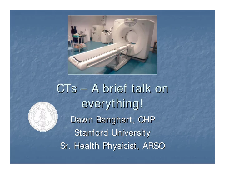
SLIDE 46

References References

  • Brenner, DJ

Brenner, DJ Computed Tomography Computed Tomography — — An Increasing Source of An Increasing Source of Radiation Exposure Radiation Exposure NEJM 2007; 357:2277 NEJM 2007; 357:2277-

  • 2284

2284

  • International Commission on Radiation Protection (ICRP)

International Commission on Radiation Protection (ICRP) Publication Publication 87 Managing Patient Dose in Computed Tomography; DEC 87 Managing Patient Dose in Computed Tomography; DEC-

  • 2001

2001

  • Brenner, DJ, Elliston C,

Brenner, DJ, Elliston C, Estimated risks of radiation Estimated risks of radiation-

  • induced fatal

induced fatal cancer from pediatric CT cancer from pediatric CT AJR AJR AJR AJR 2001;176:289 2001;176:289-

  • 296

296

  • Huda, W

Huda, W Patient radiation doses from adult and pediatric CT Patient radiation doses from adult and pediatric CT AJR AJR 2007;188(2):540 2007;188(2):540-

  • 6

6

  • Haaga

Haaga JR JR Radiation Dose Management Radiation Dose Management -

  • Weighing Risk Versus

Weighing Risk Versus Benefit Benefit AJR AJR 2001; 177:289 2001; 177:289-

  • 291

291

  • Stragegies

Stragegies for T radiation Dose Optimization for T radiation Dose Optimization

  • Lewis, MA

Lewis, MA Multislice Multislice CT: opportunities and challenges CT: opportunities and challenges BJR 74 BJR 74 (2001),779 (2001),779-

  • 781

781

  • Bushberg

Bushberg, et al., The Essential Physics of Medical Imaging, 1994 , et al., The Essential Physics of Medical Imaging, 1994

  • Mettler

Mettler, Upton, Medical Effects of Ionizing Radiation, 2008 , Upton, Medical Effects of Ionizing Radiation, 2008

slide-47
SLIDE 47

Questions? Questions?