
SLIDE 1
1
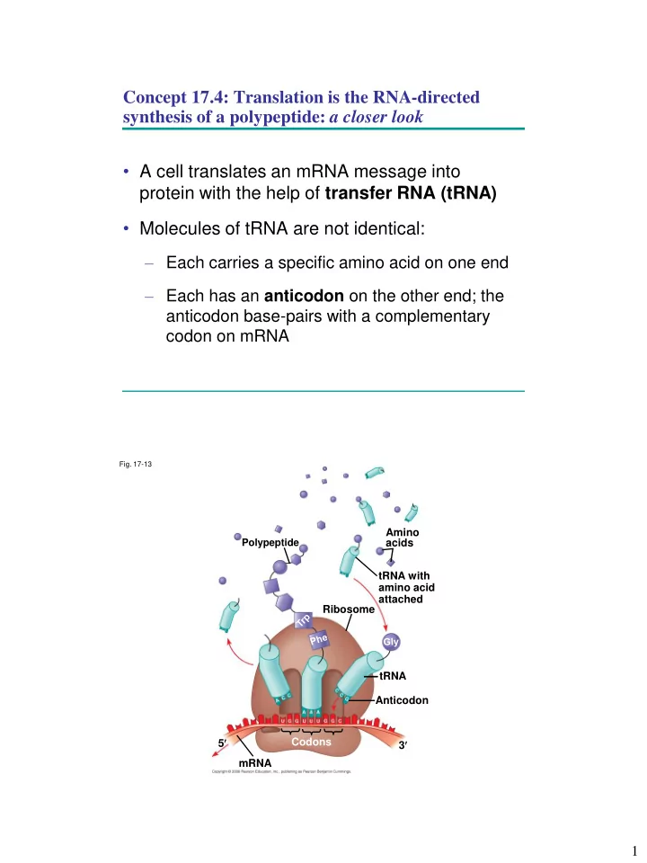
Concept 17.4: Translation is the RNA-directed synthesis of a polypeptide: a closer look
- A cell translates an mRNA message into
protein with the help of transfer RNA (tRNA)
- Molecules of tRNA are not identical:
– Each carries a specific amino acid on one end – Each has an anticodon on the other end; the anticodon base-pairs with a complementary codon on mRNA
- Fig. 17-13