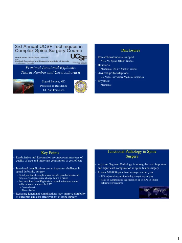

Disclosures Complications in Adult Deformity Surgery • Research/Institutional Support: – NIH, AO Spine, OREF, Globus • Honoraria: Proximal Junctional Kyphosis: – Medtronic, DePuy, Stryker, Globus Thoracolumbar and Cervicothoracic • Ownership/Stock/Options: – Co-Align, Providence Medical, Simpirica • Royalties: Sigurd Berven, MD – Medtronic Professor in Residence UC San Francisco Junctional Pathology in Spine Key Points Surgery • Readmission and Reoperation are important measures of quality of care and important contributors to cost of care • Adjacent Segment Pathology is among the most important • Junctional complications are an important challenge in and significant complication in spine fusion surgery spinal deformity surgery • In over 600,000 spine fusion surgeries per year – Distal junctional complications include pseudarthrosis and – 12% adjacent segment pathology requiring surgery progressive degenerative change below a fusion – Rates of symptomatic degeneration up to 50% in spinal – Proximal Junctional Kyphosis is related to fracture and/or deformity procedures subluxation at or above the UIV • Cervicothoracic • Thoracolumbar • Reducing junctional complications may improve durability of outcomes and cost-effectiveness of spine surgery 1
Definitions Proximal Junctional Kyphosis • Adjacent level degeneration – Radiographic signs of advanced disc degeneration or segmental instability above a fusion • Adjacent segment disease – Pathology adjacent to a fusion that creates symptoms of pain and/or nerve compression that leads to revision surgery • Proximal junctional kyphosis – Radiographic measure of greater than 5 degrees of progression of segmental kyphosis above a fusion • Proximal Junctional Failure – 10° post-operative increase in kyphosis between upper instrumented vertebra (UIV) and UIV+2, along with one or more of the following: fracture of the vertebral body of UIV or UIV +1, posterior osseo-ligamentous disruption, or pull-out of instrumentation at the UIV. • Kyphotic Decompensation Syndrome – Progressive sagittal deformity requiring revision surgery for realignment of the spine Etiology and Pathogenesis • Proximal Junctional Kyphosis – Choice of Levels • Suk S, et al: Spine 2006 – Stopping at or distal to T11 increases risk of adjacent segment kyphosis (50% PJK) – Radiographic Factors • Swank S, et al: JBJS 1981 – Fusions from L1or L2 to the sacrum have an unacceptable rate of mechanical failure – Biomechanics (7/20) • • Rigidity of Fixation Simmons ED, et al: SRS 2005 – 60% adjacent segment “topping off” in long fusions with cephalad level of L1,L2 – Patient-specific Factors • Glattes CG, et al: Spine 2005 – 26% incidence of PJK in long adult deformity constructs. Highest at T3. Little • Bone Quality impact on clinical outcome. • Age • Hostin R and ISSG: Spine 2012 – 5.6% incidence of Acute Proximal Junctional Failure (68/1218) • Neuromuscular Pathology • Defined as 15degrees proximal kyphosis, Fracture at or above UIV • Or need for revision surgery within 6 mos 2
• Defining PJK: • Restrospective study of 157 consecutive patients with • long fusion for deformity 62/161 pts with adult deformity and fusions >5 levels (39%) at 7.8yr f/u • 59% within 8 weeks • PJK observed in 32 (20%) • Risk factors: – Older age (>55yo) – Posterior instrumentation – Combined A/P surgery – Fusion to sacrum – Pedicle screws (age non-adjusted) – Significant sagittal imbalance – LIV at S1 (age non-adjusted) • Outcome worst with kyphosis >20 degrees • TK+LL+PI>45 degrees • Rate not dependent upon proximal level • SVA change more than 5cm • No association with age, BMI, BMD Proximal Junctional Kyphosis UCSF Experience: • 125 adults with proximal fusions T9-L2 Maruo K, UCSF Spine Service: Spine – 90 consecutive patients fused from T9-L1 to pelvis – Average 7.1 levels fused – Average Age- 64.5 • 3 groups sorted by PIV PJK Revision – Minimum Follow-up 2 years (2.9 years average) – T9-10 51% 24% – Radiographic PJK observed in 37 patients (41%) – T11-12 55% 24% – Reoperation in 12 patients (12%) – L1-L2 36% 26% – Purpose: • Recommendation: Choose lowest neutral and stable • Defined Risk Factors for PJK proximal vertebra • Identify Protective Strategies 3
• 68yo male physician with progressive sagittal and coronal plane deformity • Lower back pain with limited neurogenic symptoms 4
5
Proximal Junctional Kyphosis UCSF Experience: Maruo et al: Spine in Press – 90 consecutive patients fused from T9-L1 to pelvis – Radiographic PJK observed in 37 patients (41%) – Reoperation in 12 patients (12%) – Risk factors: • Change in Lumbar Lordosis >30 degrees • Pre-operative thoracic kyphosis >30 degrees • Preoperative PJA >10 degrees • Pelvic incidence >55 degrees – Protective strategy • Post-op SVA<50mm, PT<20 degrees, and PI-LL<+/-10 degrees Cervicothoracic Junctional Pathology • Upper Thoracic vs Thoracolumbar End Vertebra 6
90 38 38 39 39 90 90 7
4 weeks post-op Patient with severe cervicothoracic pain 8
9
3 year follow-up 10
10 pts with PJK 5 with UVI collapse and adjacent subuxation 5 with adjacent fx Risk factors: Osteopenia, Large sagittal plane correction, old age, comorbidities Decompensation in first 6 mos High rate 2/5 of neural compromise in pts with UVI collapse and adjacent subluxation 11
Proximal Junctional Kyphosis Proximal Junctional Kyphosis UCSF Experience: Ha Y, UCSF Spine Service: J Neurosurg Spine August, 2013 162 consecutive adults with long fusions to the sacrum 127 distal thoracic (T9 to L1) 35 proximal thoracic (T2 to T5) Radiographic PJK 31% distal thoracic 25% proximal thoracic Kyphotic decompensation disease 6.3% distal thoracic 5.7% proximal thoracic Mechanism of distal thoracic decompensation was fracture at UIV Mechanism of proximal thoracic decompensation was subluxation- 2 cases with neural injury Evidence-based Approach to Choosing a Level Indications for Extending Arthrodesis to the Upper Thoracic • Criteria for revision in PJF: Spine – 27/59 patients with PJF underwent revision surgery within • Extension of measured curve to the structural thoracic spine 6 months of the index operation • Segmental kyphosis at the thoracolumbar junction – Patients with combined posterior/anterior approaches – >5 degrees – Patients with more extreme PJK angulation • Thoracic Kyphosis >30 degrees – Patients sustaining trauma were also significantly more • Osteoporosis likely to undergo revision • Neuromuscular Disease – Upper thoracic versus thoracolumbar proximal junction did NOT influence decision for revision 12
Promising solutions? Risk Factors for PJK • Osteoporosis • Fusion to the sacrum • Decompression Only • Fate of the L5-S1 • Choice of proximal levels intervertebral disc • Supralaminar fixation • Posterior Fusion vs. Circumferential Arthrodesis • Correction of lordosis >30 degrees w/o PSO • Cephalad extent of arthrodesis • Mismatch of Lumbar Lordosis and PI • The role of iliac fixation • Pre-operative thoracic kyphosis >30 degrees • Osteoporosis – Pre-op PJA >10 degrees • Rigidity of construct? Possible solutions Vertebral Augmentation and PJK • Minimize cantilever forces at cephalad end of construct • Matching Lumbar Lordosis to Pelvic Incidence – PI+LL+TK<45 0 • Augmentation of proximal fixation • Augmentation of level above proximal fixation • Interspinous augmentation/stabilization • Dynamic stabilization 13
14
• Transitional rod at UIV results in: – Reduced nuclear pressure at adjacent disc – Reduced angular displacement of adjacent segment – Reduced strain on cephalad screw 15
Evidence-based Approach to PJK in Deformity Surgery • Match Lumbar Lordosis and Pelvic Incidence – LL+TK+PI<45 degrees • Choice of Levels – Extend to upper thoracic spine • PJA>5 degrees, TK > 30 degrees, Osteoporosis • Limit Correction – Osteoporosis, Longstanding deformity, Neuromuscular conditions • Vertebral Augmentation at and/or above UIV • Dynamic Stabilization of UIV Conclusions Thank you • Reoperations are an important measure of quality, and contributor to cost of care in adult deformity • Proximal Junctional Kyphosis is a common cause for reoperation in adult deformity • Surgical strategies to reduce junctional kyphosis may reduce the cost of care and improve quality of care UCSF Center for Outcomes Research 16
17
18
Recommend
More recommend