CNS SESSION HPI: 46 yo right handed woman who presented to her PCP - - PDF document

cns session
SMART_READER_LITE
LIVE PREVIEW

CNS SESSION HPI: 46 yo right handed woman who presented to her PCP - - PDF document

3/8/2018 18 th Multidisciplinary Management of Cancers: A Casebased Approach 18 th Multidisciplinary Management of Cancers: A Casebased Approach CASE 1 CNS SESSION HPI: 46 yo right handed woman who presented to her PCP with increasing


slide-1
SLIDE 1

3/8/2018 1

18th Multidisciplinary Management of Cancers: A Case‐based Approach

CNS SESSION

Chair: Ruben Fragoso, MD/PhD—UC Davis Fellow: Michael Cardenas, MD—UC Davis Panel: Gordon Li, MD—Stanford Seema Nagpal, MD—Stanford Jennie Taylor, MD—UCSF

18th Multidisciplinary Management of Cancers: A Case‐based Approach

HPI: 46 yo right handed woman who presented to her PCP with increasing visual changes and left hand numbness over three weeks in mid April 2017. CT of the head showed a right temporal intra‐axial mass with a 6 mm midline shift. CT of the chest/abdomen/pelvis was negative. On exam she a had left upper quadrant visual field deficit, but no motor or sensory deficits. Her cognition appeared to be intact.

CASE 1

18th Multidisciplinary Management of Cancers: A Case‐based Approach

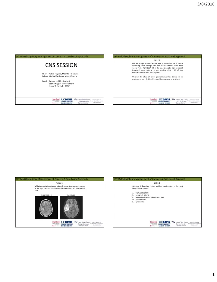
MRI at presentation showed a large 6 cm contrast enhancing mass in the right temporal lobe with mild edema and a 7 mm midline shift.

CASE 1

T1 3dFSPGR + C FLAIR CUBE

18th Multidisciplinary Management of Cancers: A Case‐based Approach

Question 1: Based on history and her imaging what is the most likely disease process? A. High grade glioma B. Low grade glioma C. Metastasis from an unknown primary D. Ependymoma E. Lymphoma

CASE 1

slide-2
SLIDE 2

3/8/2018 2

18th Multidisciplinary Management of Cancers: A Case‐based Approach

She underwent awake craniotomy with post‐operative imaging suggesting a GTR.

CASE 1

T1 FLAIR + C T1 FLAIR + C

18th Multidisciplinary Management of Cancers: A Case‐based Approach

Pathology showed WHO IV, glioblastoma, IDH 1/2 wild type. ATRX retained. TERT mutation detected. EGFRvIII and EGFR amplification negative. MGMT methylation status not reported.

CASE 1

18th Multidisciplinary Management of Cancers: A Case‐based Approach

Question 2: What adjuvant treatment would you recommend? A. 6 weeks

  • f

radiation plus concurrent and adjuvant temozolomide for 6 cycles. B. 6 weeks

  • f

radiation plus concurrent and adjuvant temozolomide for 12 cycles. C. 3 weeks

  • f

radiation plus concurrent and adjuvant temozolomide for 6 cycles. D. 3 weeks

  • f

radiation plus concurrent and adjuvant temozolomide for 12 cycles.

CASE 1

18th Multidisciplinary Management of Cancers: A Case‐based Approach

Question 2: What adjuvant treatment would you recommend?

CASE 1

Blumenthal, DT, et al., Neuro‐Oncology 2017.

slide-3
SLIDE 3

3/8/2018 3

18th Multidisciplinary Management of Cancers: A Case‐based Approach

Question 3: Alternating electric field therapy in this patient: A. Has no role in her therapy. B. Should be reserved for when she has a recurrence. C. Can be used in the adjuvant phase along with temozolomide. D. Should be used only on weekends during the six weeks of radiation.

CASE 1

18th Multidisciplinary Management of Cancers: A Case‐based Approach

Question 3: Alternating electric field therapy in this patient:

CASE 1

Stupp, R, et al., JAMA 2017.

18th Multidisciplinary Management of Cancers: A Case‐based Approach

She completed 60 Gy of radiation along with concurrent temozolomide in mid‐July 2017. She tolerated treatment well. Her post chemoradiation scan (~5wks) showed:

CASE 1

post‐surgery scan T1 + C 5 week post RT scan

18th Multidisciplinary Management of Cancers: A Case‐based Approach

Question 4: Her post radiation MRI scan shows changes that likely represent? A. Pseudoprogression. B. Tumor progression. C. Either A or B as they cannot be distinguished on MRI. D. Typical radiation induced imaging changes.

CASE 1

slide-4
SLIDE 4

3/8/2018 4

18th Multidisciplinary Management of Cancers: A Case‐based Approach

She was started on adjuvant temozolomide along with alternating electric field therapy . Her subsequent three month scan showed:

CASE 1

T1 + C 5 week scan 3 month scan

18th Multidisciplinary Management of Cancers: A Case‐based Approach

Question 5: What scan would be least helpful in differentiating between pseudoprogression and tumor progression? A. CT of the head with/without contrast. B. MR spectroscopy C. MR perfusion D. PET/BRAIN

CASE 1

18th Multidisciplinary Management of Cancers: A Case‐based Approach

The imaging studies showed significantly elevated cerebral blood volume in the area of nodular enhancement and significant interval increase in T2/FLAIR.

CASE 1

T1 + C FLAIR CUBE rCBV

18th Multidisciplinary Management of Cancers: A Case‐based Approach

Question 6: Given her MRI findings and the timing of her radiation what options does she still have left (aside from a clinical trial)? A. Surgery B. Chemotherapy C. Fractionated re‐irradiation D. Radiosurgery

CASE 1

slide-5
SLIDE 5

3/8/2018 5

18th Multidisciplinary Management of Cancers: A Case‐based Approach

Radiation and further surgery were not recommended. She continued

  • n

alternating electric field therapy and her chemotherapy was changed to bevacizumab and CCNU.

CASE 1

18th Multidisciplinary Management of Cancers: A Case‐based Approach

HPI: 50 year old right handed woman with a previous history of a right temporal meningioma initially diagnosed in 2000 now seen for a right frontal lesion. She was seen for initial consult in 2010 for Gamma Knife radiosurgery assessment for her recurrent meningioma after having surgery twice (2000 and 2007) and radiosurgery (2005). She again underwent radiosurgery in 2010 and was followed with MRI surveillance imaging which showed the progressive right frontal lesion.

CASE 2

18th Multidisciplinary Management of Cancers: A Case‐based Approach

Surveillance imaging showed a stable meningioma in the right paraclinoid region but also showed a slowly enlarging non‐contrast enhancing lesion in the medial right frontal lobe. She was asymptomatic.

CASE 2 September 2010

T1 + C T1 + C T2 FLAIR

18th Multidisciplinary Management of Cancers: A Case‐based Approach

Surveillance imaging showed a stable meningioma in the right paraclinoid region but also showed a slowly enlarging non‐contrast enhancing lesion in the medial right frontal lobe. She was asymptomatic.

CASE 2

6/2011 1/2014 12/2016

T2 FLAIR

slide-6
SLIDE 6

3/8/2018 6

18th Multidisciplinary Management of Cancers: A Case‐based Approach

Question 1: The imaging characteristics are most consistent with what type of glioma? A. JPA B. Low grade glioma C. Anaplastic glioma D. GBM

CASE 2

18th Multidisciplinary Management of Cancers: A Case‐based Approach

After tumor board discussion, craniotomy was offered and performed in June 2017. A GTR was achieved.

CASE 2

T2 FLAIR pre‐op T2 FLAIR post‐op

18th Multidisciplinary Management of Cancers: A Case‐based Approach

Pathology returned as:

  • Anaplastic diffuse glioma (oligodendroglioma phenotype), WHO grade III
  • IDH1 R132H mutation not present (IHC) and resent for PCR assessment and

found to be c.394>T (p.R132C) mutated

  • ATRX retained (IHC)
  • 1p19q NOT co‐deleted (FISH)
  • TERT promoter mutation not detected (PCR)
  • MGMT promoter methylation detected (PCR)

CASE 2

18th Multidisciplinary Management of Cancers: A Case‐based Approach

Question 2: What genetic/molecular marker finding on her pathology is most important for treatment management? A. IDH1 mutation B. Non‐codeletion of 1p19q C. MGMT methylation D. TERT promoter mutation E. None of the above

CASE 2

slide-7
SLIDE 7

3/8/2018 7

18th Multidisciplinary Management of Cancers: A Case‐based Approach

Question 3: According to the CATNON trial (EORTC study 26053‐ 22054) interim analysis optimal treatment for this patient is suggested to be? A. Radiation alone. B. Radiation and concurrent temozolomide. C. Radiation and adjuvant temozolomide. D. Radiation and concurrent plus adjuvant temozolomide.

CASE 2

18th Multidisciplinary Management of Cancers: A Case‐based Approach

The patient completed radiation with concurrent temozolomide early September 2017 with 6‐12 cycles of adjuvant temozolomide planned.

CASE 2

18th Multidisciplinary Management of Cancers: A Case‐based Approach

Question 4: Of the regimens below what is the most appropriate MRI surveillance schedule after completing radiation? A. At 6 wks, then every 3 months for 3 years, then every 6 months. B. At 6 wks, then every 6 months for 3 years, then every year. C. At 6 wks, then every 3 months for 5 years, then as clinically indicated. D. At 6 wks, then every 6 months for 5 years, then as clinically indicated.

CASE 2

18th Multidisciplinary Management of Cancers: A Case‐based Approach

HPI: 27 year old right handed woman presented with a history of low grade glioma now with evidence of recurrence. She was initially diagnosed in 2014 after a period of new headaches, intermittent vision changes, and paresthesias of the right hand, lip and tongue. A MRI was performed. The MRI at the time showed a non‐enhancing right frontal lesion consistent with glioma.

CASE 3

slide-8
SLIDE 8

3/8/2018 8

18th Multidisciplinary Management of Cancers: A Case‐based Approach

Question 1: Maximal safe resection should be attempted for which

  • f the following consideration(s):

A. Higher grade foci may not be seen in a small sample. B. Less tumor to possibly dedifferentiate to a higher grade. C. Decreased tumor load may enhance subsequent therapy. D. All of the above.

CASE 3

18th Multidisciplinary Management of Cancers: A Case‐based Approach

She underwent a craniotomy in July 2014. The tumor was debulked to the maximum extent possible. Pathology showed an infiltrating astrocytoma, WHO grade II.

CASE 3

18th Multidisciplinary Management of Cancers: A Case‐based Approach

Question 2: Which genetic alteration is most probable? A. 1p19q co‐deletion. B. IDH2 mutation. C. IDH1 mutation. D. IDH1/2 and 1p19q co‐deletion.

CASE 3

18th Multidisciplinary Management of Cancers: A Case‐based Approach

Question 2: Which genetic alteration is most probable?

CASE 3

Yan, H, et al., NEJM 2009

slide-9
SLIDE 9

3/8/2018 9

18th Multidisciplinary Management of Cancers: A Case‐based Approach

Pathology showed an infiltrating astrocytoma, WHO grade II, with IDH1 (R132H) mutation positive, p53 positive, ATRX mutated, MIB 5%, and BRAF (V600E) mutation negative. She had a wound infection and subsequently a revision for

  • dehiscence. Temozolomide was given May 2015 ‐ June 2016 (15

cycles).

CASE 3

18th Multidisciplinary Management of Cancers: A Case‐based Approach

MRI in August 2016 showed a persistent mass and now with increased blood perfusion and abnormal diffusion along the parasagittal region.

CASE 3

CUBE T2 FLAIR

18th Multidisciplinary Management of Cancers: A Case‐based Approach

Question 3: What management course is the least optimal? A. Radiation and adjuvant PCV. B. Radiation alone. C. Surgery with an attempt at GTR. D. Observation until she becomes symptomatic.

CASE 3

18th Multidisciplinary Management of Cancers: A Case‐based Approach

After tumor board discussion, she again underwent resection in September 2016. Pathology was consistent with the previous specimen.

CASE 3

Pre‐op Post‐op

slide-10
SLIDE 10

3/8/2018 10

18th Multidisciplinary Management of Cancers: A Case‐based Approach

Option of pursuing definitive radiation and chemotherapy was discussed versus pursuing at the time of progression. She was followed with serial imaging which showed evidence of slow progression in April 2017. After tumor board presentation, the consensus was to proceed with radiation and nitrosourea‐ based chemotherapy.

CASE 3

April 2017 Post‐op

18th Multidisciplinary Management of Cancers: A Case‐based Approach

Question 4: Which of the following radiation regimens is not appropriate? A. 3D conformal therapy to 54 Gy in 1.8 Gy fractions. B. IMRT to 54 Gy in 1.8 Gy fractions. C. Stereotactic radiosurgery with a marginal dose of 15 Gy. D. Whole brain radiation to 30 Gy in 3 Gy fractions. E. Both C and D are not appropriate.

CASE 3

18th Multidisciplinary Management of Cancers: A Case‐based Approach

Question 5: In retrospect, at initial diagnosis, if tumor invaded into the corpus callosum, according to RTOG 9802 she would most likely be? A. low risk and require no further therapy. B. high risk and require immediate RT and adjuvant PCV. C. high risk and require delayed RT and adjuvant PCV until progression. D. high risk but require no further therapy.

CASE 3

18th Multidisciplinary Management of Cancers: A Case‐based Approach

HPI: 67 yo man with a distant smoking history diagnosed with metastatic lung cancer in early 2015 after having a persistent worsening cough. A PETCT on 1/2015 showed a right medial apical mass, mediastinal hilar lymphadenopathy and multiple bone lesions. A bronchoscopy was positive for a moderate to poorly differentiated adenocarcinoma (CK7 and TTF1 positive). Further tests (EGFR/ALK) were pending. A MRI of the brain showed two (maybe three) brain lesions, each 2‐3 mm.

CASE 4

slide-11
SLIDE 11

3/8/2018 11

18th Multidisciplinary Management of Cancers: A Case‐based Approach

HPI cont’d: Due to disease extent, worsening symptoms and having asymptomatic brain metastases he was started

  • n

carbo/pemetrexed in early 2/2015, prior to the pending studies returning. Four to six cycles were planned to be followed by erlotinib. He was referred to radiation oncology for management of his brain metastases and seen after cycle 2.

CASE 4

18th Multidisciplinary Management of Cancers: A Case‐based Approach

Question 1: Of the treatment/radiation options available to manage his brain metastases which would be most preferred? A. whole brain radiation alone. B. radiosurgery alone. C. whole brain radiation and radiosurgery. D. Surgery. E. to continue with chemotherapy as planned.

CASE 4

18th Multidisciplinary Management of Cancers: A Case‐based Approach

He underwent stereotactic radiosurgery (SRS) mid‐March 2015 (21 Gy to the 50% isodose line).

CASE 4

left temporal right occipital

18th Multidisciplinary Management of Cancers: A Case‐based Approach

Question 2: What variable most drives dose selection for radiosurgery? A. Histology B. Individual tumor volume C. Location of tumor D. Proximity of one tumor to the next E. Total number of lesions to be treated

CASE 4

slide-12
SLIDE 12

3/8/2018 12

18th Multidisciplinary Management of Cancers: A Case‐based Approach

Question 3: In what time range is radiation necrosis most commonly seen? A. Within the first two weeks from treatment. B. Within the first six months from treatment. C. Six months to two years after treatment. D. Two years and beyond after treatment.

CASE 4

18th Multidisciplinary Management of Cancers: A Case‐based Approach

His studies eventually showed an EGFR activating mutation, exon 19 deletion. Restaging PET/CT in April 2015 showed response in the lung but progression in bone. He was switched to erlotinib. He received radiation to the left hip and right scapula for increasing bone pain. MRI at the end of April 2015 showed a stable left temporal lesion and diminished right occipital lesion. Subsequent MRIs showed resolution of the treated lesions. No new lesions were seen.

CASE 4

18th Multidisciplinary Management of Cancers: A Case‐based Approach

In early December 2015, he started a Phase I trial with pembrolizumab and afatinib for progression. In early April 2016, he was enrolled and started a new Phase I trial involving erlotinib and INC280 (capmatinib). He had a dose reduction two months later for grade 3 diarrhea.

CASE 4

18th Multidisciplinary Management of Cancers: A Case‐based Approach

He continued on trial until August 2017 at which point PET/CT showed progression of pre‐existing lesions and a brain MRI showed innumerable new metastases. Blood assessment for tumor DNA now showed T790M being positive. Osimertinib was discussed by his medical oncologist.

CASE 4

T1 + C

slide-13
SLIDE 13

3/8/2018 13

18th Multidisciplinary Management of Cancers: A Case‐based Approach

Question 4: What are reasonable options at this point to manage his brain metastases? A. Whole brain radiation. B. Radiosurgery. C. Osimertinib with close MRI surveillance. D. A and C are both reasonable. E. B and C are both reasonable. F. A and B are both reasonable.

CASE 4

18th Multidisciplinary Management of Cancers: A Case‐based Approach

Patient at the time was asymptomatic. He pursued osimertinib and deferred radiation. Follow up MRIs showed less conspicuous lesions.

CASE 4

18th Multidisciplinary Management of Cancers: A Case‐based Approach

8/2017 10/2017 1/2018

CASE 4

18th Multidisciplinary Management of Cancers: A Case‐based Approach

8/2017 10/2017 1/2018

CASE 4

slide-14
SLIDE 14

3/8/2018 14

18th Multidisciplinary Management of Cancers: A Case‐based Approach

HPI: 78 yo woman presented in early July 2017 with progressive dizziness, confusion, memory loss and falls. MRI of the brain showed a heterogeneously enhancing cystic 6 cm mass in the right temporal lobe with associated T2/FLAIR hyperintensity and ependymal involvement of the right occipital horn and atrium of the lateral ventricle. There was an 8 mm midline shift. CT of the chest/abdomen/pelvis was negative.

CASE 5

T1 + C

18th Multidisciplinary Management of Cancers: A Case‐based Approach

She underwent resection with post‐operative MRI report indicating a gross total resection. Pathology was consistent with glioblastoma, WHO IV (IDH1 not mutated; MGMT methylated).

CASE 5

T1 + C

18th Multidisciplinary Management of Cancers: A Case‐based Approach

Question 1: what imaging characteristic of the tumor might be most predictive of non‐contiguous recurrence? A. Extent of heterogeneous enhancing solid component B. Overall size of tumor C. Ependymal involvement D. Extent of associated T2/FLAIR

CASE 5

18th Multidisciplinary Management of Cancers: A Case‐based Approach

Question 2: What patient factor may influence radiation recommendations? A. Age B. Extent of resection C. MGMT promoter methylation status D. IDH1 status E. None of the above

CASE 5

slide-15
SLIDE 15

3/8/2018 15

18th Multidisciplinary Management of Cancers: A Case‐based Approach

Question 3: What adjuvant treatment would you offer? A. 6 weeks RT alone B. 6 weeks RT with concurrent and adjuvant temozolomide C. 3 weeks RT alone D. 3 weeks RT with concurrent and adjuvant temozolomide

CASE 5

18th Multidisciplinary Management of Cancers: A Case‐based Approach

Question 3: What adjuvant treatment would you offer?

CASE 5

Perry, JR, et al., NEJM 2017.

18th Multidisciplinary Management of Cancers: A Case‐based Approach

She went on to receive a three week course of radiation along with temozolomide 4 weeks after craniotomy. Alternating electric field therapy was offered but she declined. Her follow up MRI six months out continues to show no evidence of disease progression.

CASE 5

T1 + C