Chapter Nine Nucleic Acids: How Structure Conveys Information Paul - - PDF document

chapter nine nucleic acids how structure conveys
SMART_READER_LITE
LIVE PREVIEW

Chapter Nine Nucleic Acids: How Structure Conveys Information Paul - - PDF document

Mary K. Campbell Shawn O. Farrell Chapter Nine Nucleic Acids: How Structure Conveys Information Paul D. Adams University of Arkansas 1 Information Transfer in


slide-1
SLIDE 1

Mary K. Campbell Shawn O. Farrell

  • Chapter Nine

Nucleic Acids: How Structure Conveys Information

Paul D. Adams • University of Arkansas

1

Information Transfer in Cells
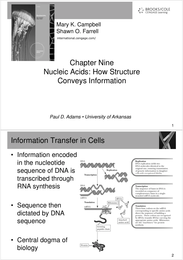
  • Information encoded

in the nucleotide sequence of DNA is sequence of DNA is transcribed through RNA synthesis

  • Sequence then

dictated by DNA dictated by DNA sequence

  • Central dogma of

biology

2

slide-2
SLIDE 2

Nucleic Acids

  • Levels of structure
  • 1°structure: the order of bases on the

polynucleotide sequence; the order of bases polynucleotide sequence; the order of bases specifies the genetic code

  • 2°structure: the three-dimensional conformation
  • f the polynucleotide backbone
  • 3°structure: supercoiling
  • 4°structure: interaction between DNA and
  • 4°structure: interaction between DNA and

proteins

3

Nucleic Acids

  • Nucleic acid:

Nucleic acid: a biopolymer containing three types of monomer units

  • a base derived from purine or pyrimidine (nucleobases)
  • a monosaccharide, either D-ribose or 2-deoxy-D-ribose
  • phosphoric acid
  • RNA (Ribonucleic Acid)
  • DNA (Deoxyribonucleic Acid)

4

slide-3
SLIDE 3

Pyrimidine/Purine Bases

  • The structures of pyrimidine and purine are shown

here for comparison

5

Other Bases

  • Less common bases can
  • ccur
  • Principally but not

exclusively, in transfer RNAs

6

slide-4
SLIDE 4

Nucleosides

  • Nucleoside:

Nucleoside: a compound that consists of D-ribose

  • r 2-deoxy-D-ribose covalently bonded to a

nucleobase by a β-N-glycosidic bond nucleobase by a β-N-glycosidic bond

  • Lacks phosphate group

7

Nucleotides

  • Nucleotide:

Nucleotide: a nucleoside in which a molecule of phosphoric acid is esterified with an -OH of esterified with an -OH of the monosaccharide, most commonly either the 3’-OH

  • r the 5’-OH
  • Name based on parent

nucleoside with a suffix “monophosphate”

  • Polymerization leads to
  • Polymerization leads to

nucleic acids. Linkage is repeated (3’,5’- phosphodiester bond)

  • (Biochemical Connections p.

232)

8

slide-5
SLIDE 5

DNA - 1° Structure

  • Deoxyribonucleic acids, DNA:

Deoxyribonucleic acids, DNA: a biopolymer that consists of a backbone of alternating units of 2-deoxy-D-ribose and phosphate

  • the 3’-OH of one 2-deoxy-D-ribose is joined to the 5’-
  • the 3’-OH of one 2-deoxy-D-ribose is joined to the 5’-

OH of the next 2-deoxy-D-ribose by a phosphodiester bond

  • Primary Structure:

Primary Structure: the sequence of bases along the pentose-phosphodiester backbone of a DNA molecule

  • base sequence is read from the 5’ end to the 3’ end
  • base sequence is read from the 5’ end to the 3’ end
  • System of notation single letter (A,G,C,U and T)

9

DNA differs from RNA

Sugar is 2’-deoxyribose, not ribose.

  • Sometimes “d” used to
  • Sometimes “d” used to

designate “deoxy”

  • Writing a DNA strand

– an abbreviated notation – even more abbreviated notations d(GACAT) d(GACAT) pdApdCpdGpdT pdACGT

10

slide-6
SLIDE 6

DNA - 2° Structure

  • Secondary structure:

Secondary structure: the

  • rdered arrangement of

nucleic acid strands

  • the double helix model of
  • the double helix model of

DNA 2°structure was proposed by James Watson and Francis Crick in 1953

  • Double helix:

Double helix: a type of 2° structure of DNA molecules in which two antiparallel in which two antiparallel polynucleotide strands are coiled in a right-handed manner about the same axis

  • structure based on X-Ray

crystallography

11 1. DNA forms a regular right-hand helix, making a complete turn every 3.4 nm with a diameter of 2 nm giving 10 nucleotides

  • nm giving 10 nucleotides

per turn. 2. The helix contains 2 anti- parallel polynucleotide chains – bases facing inwards, with a purine always

  • pposite

a pyrimidine. 3. Proportion of G always the same as the proportion of C and the proportion of T the same as A. Chargaff's Law: A=T, G=C 12

slide-7
SLIDE 7

T-A Base Pairing

  • Base pairing is complimentary
  • A major factor stabilizing the double helix is base pairing by

hydrogen bonding between T-A and between C-G

  • T-A base pair comprised of 2 hydrogen bonds

13

G-C Base Pair

  • G-C base pair comprised of 3 hydrogen bonds

14

slide-8
SLIDE 8

Other Forms of DNA

  • B-DNA

DNA

  • considered the physiological form
  • a right-handed helix, diameter 11Å
  • a right-handed helix, diameter 11Å
  • 10 base pairs per turn (34Å) of the helix
  • A-DNA

DNA

  • a right-handed helix, but thicker than B-DNA
  • 11 base pairs per turn of the helix
  • has not been found in vivo
  • has not been found in vivo
  • Z-DNA

DNA

  • a left-handed double helix
  • may play a role in gene expression

15

Comparison of A,B, and Z forms of DNA

  • Both A and B-DNA are

right-handed helices

  • Z-DNA is left handed
  • Z-DNA is left handed
  • Z-DNA occurs in nature,

usually consists of alternating purine- pyrimidine bases

  • Methylated cytosine

found also in Z-DNA

16

slide-9
SLIDE 9

Other Features of DNA

  • Base stacking
  • bases are hydrophobic and interact by hydrophobic

interactions interactions

  • in standard B-DNA, each base rotated by 32°

compared to the next and, while this is perfect for maximum base pairing, it is not optimal for maximum

  • verlap of bases; in addition, bases exposed to the

minor groove come in contact with water

  • many bases adopt a propeller-twist in which base
  • many bases adopt a propeller-twist in which base

pairing distances are less optimal but base stacking is more optimal and water is eliminated from minor groove contacts

17

Z-form is derivative of B-form

  • Produced by flipping
  • ne side of the

backbone 180˚ without backbone 180˚ without disturbing the backbone covalent bonds or hydrogen bonds

18

slide-10
SLIDE 10

Propeller Twists

  • Bases that are exposed to minor groove contact with water
  • They twist in a “propeller twist” fashion
  • Results in:
  • less optimal base pair distance
  • less optimal base pair distance
  • more optimal base pair stacking (eliminates presence of water

molecules)

19

DNA - 3° Structure

  • Tertiary structure:

Tertiary structure: the three-dimensional arrangement of all atoms of a nucleic acid; commonly referred to as supercoiling

  • Circular DNA:

Circular DNA: a type of double-stranded DNA in which the 5’ and 3’ ends of each stand are joined by phosphodiester and 3’ ends of each stand are joined by phosphodiester bonds

  • Supercoiling- Further coiling and twisting of DNA helix.
  • Topoisomerases
  • Class I: cut the phosphodiester backbone of one strand,

pass the end through, and reseal pass the end through, and reseal

  • Class II: cut both strands, pass some of the remaining

DNA helix between the cut strands, and reseal

  • DNA gyrase: a bacterial topoisomerase

20

slide-11
SLIDE 11

Super DNA Coiled Topology

  • Prokaryotic DNA is circular. It can form supercoils.
  • Double helix can be considered to a 2-stranded,

right handed coiled rope right handed coiled rope

  • Can undergo positive/negative supercoiling

21

  • 22
slide-12
SLIDE 12

Chromatin

  • The structure of chromatin
  • Each “Bead” is a

nucleosome

  • Nucleosome consists of:

DNA wrapped around histone core

  • Recent research has shown

that structure and spacing

  • f nucleosomes is

important in chromatin function.

23

Supercoiling in Eukaryotic DNA

  • Histone

Histone: a protein, particularly rich in the basic amino acids Lys and Arg; found associated with eukaryotic DNA eukaryotic DNA

  • five main types: H1, H2A, H2B, H3, H4
  • Chromatin:

Chromatin: DNA molecules wound around particles

  • f histones in a beadlike structure
  • Topological changes induced by supercoiling

accommodated by histone-protein component of chromatin.

24

slide-13
SLIDE 13

Denaturation of DNA

  • Double helix unwinds when DNA is denatured
  • Can be re-formed with slow cooling and annealing
  • Can be re-formed with slow cooling and annealing

25

Denaturation of DNA

  • Denaturation

Denaturation: disruption of 2° structure

  • most commonly by heat

denaturation (melting) denaturation (melting)

  • as strands separate, absorbance

at 260 nm increases

  • increase is called hyperchromicity
  • midpoint of transition (melting)

curve = Tm

  • the higher the % G-C, the higher
  • the higher the % G-C, the higher

the Tm

  • renaturation is possible on slow

cooling

26

slide-14
SLIDE 14

27

Principal Kinds of RNA

  • RNA
  • consist of long, unbranched chains of nucleotides

joined by phosphodiester bonds between the 3’-OH of joined by phosphodiester bonds between the 3’-OH of

  • ne pentose and the 5’-OH of the next
  • the pentose unit is β-D-ribose (it is 2-deoxy-D-ribose

in DNA)

  • the pyrimidine bases are uracil and cytosine (they are

thymine and cytosine in DNA)

  • in general, RNA is single stranded (DNA is double

stranded)

28

slide-15
SLIDE 15

29

RNA

  • RNA molecules are classified according to their

structure and function

30

slide-16
SLIDE 16

tRNA

  • Transfer RNA, tRNA:

Transfer RNA, tRNA:

  • the smallest kind of the

three RNAs three RNAs

  • a single-stranded

polynucleotide chain between 73-94 nucleotide residues

  • carries an amino acid at

its 3’ end its 3’ end

  • intramolecular hydrogen

bonding occurs in tRNA

31 32

slide-17
SLIDE 17

rRNA

  • Ribosomal RNA,

Ribosomal RNA, rRNA rRNA: a ribonucleic acid found in ribosomes, the site of protein synthesis

  • only a few types of rRNA exist in cells
  • only a few types of rRNA exist in cells
  • ribosomes consist of 60 to 65% rRNA and 35 to 40%

protein

  • in both prokaryotes and eukaryotes, ribosomes

consist of two subunits, one larger than the other

  • analyzed by analytical ultracentrifugation
  • analyzed by analytical ultracentrifugation
  • particles characterized by sedimentation coefficients,

expressed in Svedberg units (S)

33

The Structure of the Prokaryotic Ribosome

34

slide-18
SLIDE 18
  • 35

mRNA

  • Messenger RNA, mRNA

Messenger RNA, mRNA: a ribonucleic acid that carries coded genetic information from DNA to ribosomes for the synthesis of proteins ribosomes for the synthesis of proteins

  • present in cells in relatively small amounts and very

short-lived

  • single stranded
  • biosynthesis is directed by information encoded on

DNA

  • a complementary strand of mRNA is synthesized

along one strand of an unwound DNA, starting from the 3’ end

36

slide-19
SLIDE 19

snRNA

  • Small nuclear RNA (snRNA) is a recently

discovered RNA

  • Found in nucleus of eukaryotes
  • Small (100-200 nucleotides long)
  • Forms complexes with protein and form small
  • Forms complexes with protein and form small

nuclear ribonucleoprotein particles (snRNPs)

  • snRNPs help with processing of initial mRNA

transcribed from DNA

37