SLIDE 1
Cellular Adaptation
SLIDE 2
The very best way for most minds to remember, or identify, or understand a disease is to associate it with a morphologic IMAGE. This can be gross, electron microscopic, light microscopic, radiologic, or molecular. In MOST cases it is at the LIGHT MICROSCOPIC LEVEL.
SLIDE 3 The cell is the fundamental unit of disease
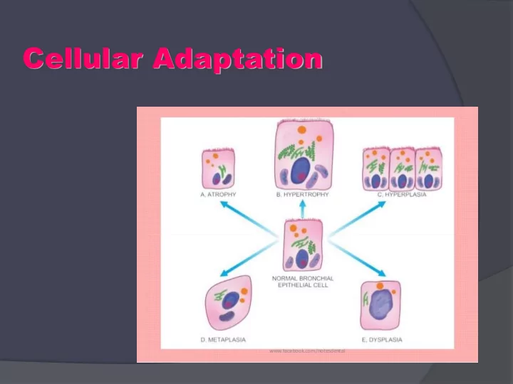
SLIDE 4 CELLULAR ADAPTATION: The ability of cells to respond to various types of stimuli and adverse environmental changes.
The cell changes chat occur are:
- Atrophy(reduction in size and cell number)
Hypertrophy (enlargement of individual cells)
- Hyperplasia, (increase in cell number)
- Metaplasia(transformation from one type
- f epithelium to another)
- Dysplasia(disordered growth of cells)
- Anaplasia,,,, Neoplasia.
SLIDE 5 Cellular Changes
Then anaplasia/neoplasia
SLIDE 6
HYPERPLASIA
Basic description: Increase in the number of cells. Types of hyperplasia Physiologic hyperplasia: Occurs due to a normal stressor. For example, increase in the size of the breasts during pregnancy, increase in thickness of endometrium during menstrual cycle, and liver growth after partial resection. Pathologic hyperplasia: Occurs due to an abnormal stressor. proliferation of endometrium due to prolonged estrogen stimulus. Important point regarding hyperplasia: Only cells that can divide will undergo hyperplasia; therefore, hyperplasia of the myocytes in the heart and neurons in the brain does not occur.
SLIDE 7
1- FOL LICULAR HYPERPLASIA (LYMPH NODE) 2- FOLLICULAR HYPERLASIA (TONSIL) 3- SINUS HYPERPLASIA (LYMPH NODE)
SLIDE 8
LYMP MPH H NODE DE
SLIDE 9
SLIDE 10
SLIDE 11
- FOL LICULAR HYPERPLASIA (LYMPH NODE)
- ةفلتخم موجحب ةبرجلؤا نوكتو ،اهمجحو ،ةبرجلؤا ددع ةدايز.
- بارجلا مظعم لمشيل جوتنلا زكرم ةماخض.
- ةبرجلؤا مّخضتل ةجيتن ةظفحملا تحت بويجلا لوزت.
- ،بللا ىّتح لصتو رشقلا زواجتت ثيح ةدقعلا مظعم ةبرجلؤا حاتجت
ةظفحملا زواجتت لب اهّنكلو.
SLIDE 12
SLIDE 13
SLIDE 14
SLIDE 15
SLIDE 16
SLIDE 17 2- FOL OLLICULAR ICULAR HYP YPERLASIA ERLASIA (TON ONSIL) SIL)
ةزوللا يف يبارج عنصت طرف
ةدقعلا فلبخب كلذو ،ةزوللا يّطغت ةنّرقتم ريغ ةيطاخم ةقّبطم ةيفصر ٌةرشب يفيللا جيسنلا نم ةظفحم اهب طيحت يتلا ةيوافمللا(ةزيمم ةملبع يهو ،) ةيوناثو ةيلوأ ًاراوغأ ًةنّوكم قمعلل حطسلا نم تادامغنا ةرشبلا هذه يدبت جوتن زكرم تاذو ،ريبك اهمجح عّنصتلا ةطرفم ةيفمللا ةبرجلؤا ىرن ةرشبلا تحت مخض ةيباعل ددغ نم ءزج رّضحملا يف ىرن نأ نكمملا نم
SLIDE 19
SLIDE 20
SINUS HYPERPLASIA (LYMPH NODE)
ةيفمللا ةدقعلا يف يبيج عّنصت طرف
ىلإ يّدؤي كلذو ةيبللا ًاصوصخو ةيفمللا ةدقعلا يف بويجلا عّسوت ظحلبنو ةبرجلؤا رمضت ام ًابلاغ كلذلو رشقلا باسح ىلع بللا مخضت اهضعبل اياقب . ةظفحملا لوح محشلاو ةيفيللا ةظفحملا. رشقلا : ،لب وأ ةرماض نوكت دق يتلا ةيفمللا ةبرجلؤا ضعب هيف دهاشي ًاضيأ مخضتت امبرو. بللا :علابلا ايلبخلاب ئلتمتو ةيفمللاو ةيديرولا بويجلا عّسوتت ثيح ة ةيمزلببلاو ةيفمللاو
SLIDE 21
SLIDE 22
SLIDE 23
SLIDE 24
SLIDE 25
HYPERTROPHY
Increase in the size of the cell. Types of hypertrophy Physiologic hypertrophy: Occurs due to a normal stressor. For example, enlargement of skeletal muscle with exercise. Pathologic hypertrophy: Occurs due to an abnormal stressor. For example, increase in the size of the heart due to aortic stenosis. Morphology of hyperplasia and hypertrophy: Both hyperplasia and hypertrophy result in an increase in organ size; therefore, both cannot always be distinguished grossly, and microscopic examination is required to distinguish the two
SLIDE 26 ATROPHY
- Atrophy is the shrinkage in cell size by loss of cellular
substance
- With the involvement of a sufficient number of cells, an entire
- rgan can become atrophic
- Causes of atrophy include decreased workload, pressure,
diminished blood supply or nutrition, loss of endocrine stimulation, and aging
- Mechanisms of atrophy : Apoptosis..Autophagy..
SLIDE 27
1- Fatty changes,,fatty degenration,,,lymph node.
2- Fatty changes …degenration…thymus.
SLIDE 28
ةيفمللا ةدقعلا يف يمحش عجارت
ةظفحملا جراخ يأ ةدقعلا جراخ ةيمحش ايلبخ ظحلبن نأ يعيبطلا نم ةيفيللا (يوس لكشب ) ةحضاو ةيمحش ايلبخ ترهظ لاح يف نكلو ثودح ىلع لدي اذهف ةيفمللا ةدقعلل يولخلا ميشناربلا نمض دودحلا ةيفمللا ةدقعلا هذه يف يمحش عجارت ةرماضلا ةيفمللا ةبرجلؤا ضعب هيف رشق. ةيفمللا ةدقعلا وزغي يمحش جيسن.
SLIDE 29
SLIDE 30
SLIDE 31
SLIDE 32 Histologic Features of the Thymus The cortex is composed primarily of lymphocytes(thymocytes), with a few epithelial and mesenchymal Cells , whereas the medulla is composed of more epithelial cells but fewer
- lymphocytes. Hassall corpuscles are the characteristic, round,
keratinized formations with mature epithelial cells.
SLIDE 33
SLIDE 34
سومياتلا يف يمحش عجارت جيسنلا اياقبو يمحش جيسنو ماض جيسن يفمللاو يورشبلا. يهو بللا يف ةدوجوم لاساه تاميسج اياقب حئافص نم ةنّوكم ةنّرقتم ةيولخ ريغ ىنب ربتعيو ،ينيزويأ نولب رهظت زكرملا ةدحّتم ةمّلبعلا ةطقنلا لاساه ميسج
SLIDE 35
SLIDE 36
SLIDE 37 METAPLASIA
REVERSIBLE change in which one mature/adult cell type (epithelial or mesenchymal) is replaced by another mature cell type A protective mechanism rather than a premalignant change
Physiological metaplasia: cervical ectopy Pathological metaplasia Occurs as a response to chronic chemical or physical stimuli May completely regress or lead to malignant transformation (considered precancerous) Examples Intestinal metaplasia (Barrett metaplasia) Squamous metaplasia of the bronchi due to smoking → ciliated pseudostratified columnar epithelium is replaced by stratified squamous epithelium Squamous metaplasia of the bladder due to Schistosoma infection, urinary calculi, or indwelling catheters
SLIDE 38
SLIDE 39 Ciliated columnar cells Stratifiied squamous cells
SLIDE 40
SLIDE 41
SLIDE 42 1 . ةيسفنت ةراهظ. .2ةيكيبلام ةراهظ. .3يوحت ةيطاخم تحت ةقبط ددغو وخر ماض جيسن. .4يفورضغ جيسن
SLIDE 43
SLIDE 44
SLIDE 45
SLIDE 46 Barrett’s Esophagus Intestinal Metaplasia of the Esophagus
SLIDE 47
SLIDE 48
SLIDE 49
THANK YOU