Case Report Disappearing Brugada Pattern : A Rare Presentation 1 2 - - PDF document

case report
SMART_READER_LITE
LIVE PREVIEW

Case Report Disappearing Brugada Pattern : A Rare Presentation 1 2 - - PDF document

Vidarbha Journal of Internal Medicine Volume 27 July 2019 Case Report Disappearing Brugada Pattern : A Rare Presentation 1 2 3 4 Anil R Jawahirani , Mahesh Fulwani , Sachin Jibhkate , Raviraj Pardesi ABSTRACT A 60 years old lady


slide-1
SLIDE 1

VJIM Volume 27 July 2019 108 Vidarbha Journal of Internal Medicine Volume 27 July 2019

Disappearing Brugada Pattern : A Rare Presentation

Case Report

1 2 3 4

Anil R Jawahirani , Mahesh Fulwani , Sachin Jibhkate , Raviraj Pardesi Introduction : Brugada syndrome is a distinct arrhythmogenic disorder widely recognized as a sudden cause of death in the young. It is identified by a classical ST segment elevation on electrocardiogram (ECG) that may be provoked in the context of a fever or vagal

  • stimulation. The pathophysiology and genetic basis

have been elucidated as an abnormality in ion

  • channels. The management is centered around

device therapy with the implantable cardioverter defibrillator (ICD), though pharmacological treatments are being actively pursued. Case Report : A 60 years old lady with no significant past medical history presented in April 2019 with chief complaints of fever with chills and body ache since 7

  • days. She had trauma to right foot followed by

swelling, redness and fever since 7 days. On examination she had tachycardia, fever (temperature of 101 F), BP = 110/70 mm Hg, respiratory rate of 30/min, SPO2 of 98% at room air and icterus. Local examination revealed right lower

ABSTRACT A 60 years old lady with no significant past medical history presented to our hospital with cellulitis right lower limb. She was in sepsis with multi organ dysfunction. On work up she had a WBC count of 57,300/cmm, with 95%

  • polymorphs. Her hepatic and renal parameters were deranged. Her ECG was suggestive of Type I Brugada syndrome,

Echocardiography was within normal limits. The patient was treated with higher antibiotics (Piperacillin / Tazobactum with Linezolid), chymoral forte and Mg sulphate dressing. Her fever and systemic dysfunction gradually

  • improved. Since she had no cardiac symptoms, the ECG was done on alternate days. The Brugada manifestations

gradually reduced and disappeared on 8th day. She was discharged on 14th day with oral antibiotics and dressing of right lower limb with normal ECG. She is kept in close follow up for fever induced Brugada syndrome.

  • 1Asst. Prof., Department of Cardiology, JNMC, Sawangi, Wardha

2Director and Interventional Cardiologist, Shree Krishna

Hrudayalaya & Critical Care Center, Nagpur

3Consultant Physician, Lata Mangeshkar Hospital, Nagpur 4Chief, Research Unit, Shree Krishna Hrudayalaya & Critical Care

Center, Nagpur Address for Correspondence - Dr. Anil Jawahirani E-mail : anilramesh123@rediffmail.com

Accepted on 19th June 2019 Received on 3rd May 2019

limb cellulitis below knee, systemic examination of CNS, RS, CVS were within normal limits and a b d o m i n a l e x a m i n a t i o n r e v e a l e d

  • hepatosplenomegaly. Her right lower limb had

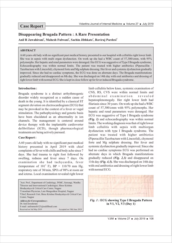
filariasis since 30 years. On work up she had a WBC count of 57,300/cmm with 95% polymorphs. Her hepatic and renal parameters were deranged. Her ECG was suggestive of Type I Brugada syndrome (Fig. 1) and echocardiography was within normal

  • limits. The working diagnosis was that of right lower

limb cellulitis with sepsis with multiorgan dysfunction with type I Brugada syndrome. The patient was treated with higher antibiotics (Piperacillin Tazobactum with Linezolid), chymoral forte and Mg sulphate dressing. Her fever and systemic dysfunction gradually improved. Since she had no cardiac symptoms ECG was performed on alternate days in which Brugada manifestations gradually reduced (Fig. 2,3) and disappeared on 11th day (Fig. 4,5). She was discharged on 14th day with oral antibiotics and dressing of right lower limb with normal ECG.

  • Fig. 1 : ECG showing Type I Brugada Pattern

in V1, V2, V3 (Day 1)

slide-2
SLIDE 2

VJIM Volume 27 July 2019 109 Vidarbha Journal of Internal Medicine Volume 27 July 2019

suggestive, but not diagnostic, of this disease (as shown in Fig. 6). Moreover, in a given patient with Brugada syndrome there are marked day-to-day changes in ECG morphology. In a large series of patients with documented Brugada syndrome who

2

underwent repeated ECG recording over the years,

  • nly every third ECG was diagnostic (i.e, showed

the type I pattern) and every third ECG was completely normal, making the diagnosis of Brugada syndrome challenging. The diagnostic criteria of Brugada syndrome are

3

described in the Table No. 1. Discussion : Brugada syndrome is a genetic disease presenting with a characteristic electrocardiogram (ECG) and a tendency to develop malignant polymorphic ventricular arrhythmias that may lead to syncope or cardiac arrest. The characteristic ECG, which includes a coved-type ST-segment elevation > 2 mm in the right precordial leads, is termed “type I” Brugada ECG pattern and is required to make the diagnosis of Brugada syndrome.1 Too often, however, the ECGs of patients with Brugada syndrome have lesser degrees or different contours

  • f ST-segment elevation (“saddleback” rather than

“coved”), which are termed type II or type III Brugada pattern. These ECG patterns are

  • Fig. 2 : ECG showing Type I Brugada Pattern

in V1, V2 (Day 3)

  • Fig. 3 : ECG showing Type I Brugada Pattern

in V1, V2, V3 (Day 5)

  • Fig. 4 : ECG showing sinus tachycardia

without any features of Brugada (Day 8)

  • Fig. 5 : ECG showing sinus tachycardia

with no features of Brugada (Day 11)

  • Fig. 6 : ECG pattern of various types of

Brugada syndrome. Table No. 1 : Diagnostic criteria of Brugada syndrome

slide-3
SLIDE 3

VJIM Volume 27 July 2019 110 Vidarbha Journal of Internal Medicine Volume 27 July 2019

useful in risk stratifying asymptomatic patients. An ICD should then be implanted where VF is inducible in the context of an incidental type-1 ECG. Alternatively, an ICD is appropriate where a sodium-channel blocker-induced ECG is precipitated in a subject who has a positive family history of sudden death. Those patients who have no family history and are symptom free but develop a type-1 ECG only after drug challenge should be kept

7

under tight surveillance. The flowchart in Fig. 7

3

serves as a useful algorithm for management. In our case since patient did not have any cardiac symptoms and her ECG at the time of discharge was normal, she is kept in close follow up. Figure 7 : Flow chart for management of Brugada syndrome Conclusion : Brugada syndrome is an inherited sodium channelopathy of cardiac myocytes. It is a major cause of sudden death in young people who have structurally normal hearts. Electrocardiography (ECG) is the investigation from which Bruagada patients are identified, though clinical features and modulating factors have a role to play. The implantable cardioverter-defibrillator is the single most effective and proven treatment in controlling Fever-induced Brugada is the term used to describe the aggravation of clinical and/or ECG characteristics of this syndrome during febrile states in susceptible individuals. The prevalence of type I Brugada ECG in patients with fever is 20 times higher than in afebrile patients, emphasizing the potency of fever in uncovering this ECG

  • phenomenon. These findings also may imply that

the number of asymptomatic Brugada patients diagnosed today is only the tip of the iceberg, as many more would have been discovered if their ECGs were recorded during febrile illnesses. All patients with fever-induced type I Brugada pattern were asymptomatic and remained so during 30

4 5

months of follow-up. In 1999, Dumaine et al reported the results of functional expression studies

  • f a genetic mutation (T1620M) identified in

patients with Brugada syndrome, showing that the loss of function of sodium channel current was accentuated at higher temperatures. The authors suggested the possibility that a febrile state may unmask the Brugada syndrome. Indeed, fever was the precipitating factor of arrhythmias in 18% of patients presenting with cardiac arrest in a large series of patients with symptomatic Brugada

6

  • syndrome. Thus, the concept of “fever-induced

Brugada syndrome” is well accepted. In our case, patient had ECG features of Type I Brugada syndrome on day 1 when she had fever with sepsis. As gradually she responded to antiobiotics, fever subsided and so the features of Brugada. Hence our case is one of the rare type of fever induced Type I Brugada syndrome. Treatment for Brugada syndrome is essentially aimed at correcting potentially life-threatening ventricular arrhythmias as they occur. There is no way of preventing arrhythmias from occurring in the first place. The single most effective and proven treatment is the ICD. Patients are electrically shocked, i.e. cardioverted into a normal sinus rhythm as a life-saving measure. These devices are the mainstay of therapy and constantly monitor the heart rhythm. Symptomatic Brugada patients should always be offered an ICD. This recommendation was borne out of a consensus conference held in

7

September 2003. Electrophysiology studies are

slide-4
SLIDE 4

VJIM Volume 27 July 2019 111 Vidarbha Journal of Internal Medicine Volume 27 July 2019

4. Fever-induced Brugada pattern: How common is it and what does it mean? Arnon Adler, MD,* Guy Topaz, BMedSc,* Karin Heller, MD,† David Zeltser, MD,‡ Tami Ohayon, MD,† Uri Rozovski, MD,§ Amir Halkin, MD,* Raphael Rosso, MD,* Shay Ben- Shachar, MD,¶Charles Antzelevitch, PhD, FHRS,? and Sami Viskin, MD* 2013 Jul 19. doi: 10.1016/j.hrthm.2013.07.030 5. Dumaine R, Towbin JA, Brugada P, et al. Ionic mechanisms responsible for the electrocardiographic phenotype of the Brugada syndrome are temperature dependent. Circ Res. 1999;85:803-809. [PubMed] [Google Scholar] 6. Amin AS, Meregalli PG, Bardai A, Wilde AA, Tan HL. Fever increases the risk for cardiac arrest in the Brugada syndrome. Ann Intern Med. 2008;149:216-218. [PubMed] [Google Scholar] 7. Schulze-Bahr E, Eckardt L, Breithardt G et al. Sodium channel gene (SCN5A) mutations in 44 index patients with Brugada syndrome: different incidences in familial and sporadic disease. Hum Mutat2003;2:651-2. Https://doi.org/10.1002/humu.9144.

life-threatening ventricular arrhythmias. Fever is an important trigger and the onset of pyrexia (such as that seen in our patient) may unmask abnormal ECG morphology and raise suspicion of Brugada syndrome. References :

1. Antzelevitch C, Brugada P, Borggrefe M, et al. Brugada syndrome: report of the second consensus conference: endorsed by the Heart Rhythm Society and the European Heart Rhythm Association.

  • Circulation. 2005;111:659-670. [PubMed] [Google Scholar]

2. Richter S, Sarkozy A, Veltmann C, et al. Variability of the diagnostic ECG pattern in an ICD patient population with Brugada

  • syndrome. J Cardiovasc Electrophysiol. 2009;20:69-75. [PubMed]

[Google Scholar] 3. Benito B, Brugada R, Brugada J, Brugada P. Brugada syndrome. ProgCardiovasc Dis 2008;51:1-22. Https://doi.org/10.1016/j.pcad. 2008.05.002