Case presentation History Abandoned at the age of 1,5 y, since - - PDF document

case presentation
SMART_READER_LITE
LIVE PREVIEW

Case presentation History Abandoned at the age of 1,5 y, since - - PDF document

4/18/2018 Center for Congenital Heart Diseases Center for Congenital Heart Diseases July 2006 Case presentation History Abandoned at the age of 1,5 y, since then in a children's home where he 15 yo boy with PAH/CHD stayed at 3000 m


slide-1
SLIDE 1

4/18/2018 1

Center for Congenital Heart Diseases

University Medical Center Groningen

Theresia Vissia-Kazemier, RN MANP

Dutch National Network for Pediatric Pulmonary Hypertension Center for Congenital Heart Diseases

Department of Pediatric Cardiology Beatrix Children’s Hospital, UMCG

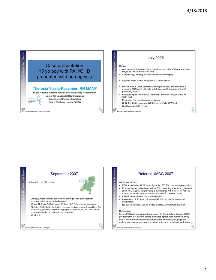
Case presentation 15 yo boy with PAH/CHD presented with hemoptysis

Center for Congenital Heart Diseases

University Medical Center Groningen

July 2006

History

  • Abandoned at the age of 1,5 y, since then in a children's home where he

stayed at 3000 m altitude in China

  • Cyanotic boy, “nothing could be done for him in Beijing”
  • Adopted from China at the age of 4 y, Dutch family
  • Presentation at Dutch pediatric cardiologist (Leiden) who confirmed a

moderate ASD type II with right to left shunt and hypoplasia of the left pulmonary artery

  • Echocardiogram: RVP about 140 mmHg, moderate function of the RV,

WHO fc III

  • Medication: furosemide en spironolacton
  • RHC: Tcsat 86%, systemic RVP 80 mmHg, PVRi 11 WU.m2
  • Start bosentan tid 31¼ mg

Center for Congenital Heart Diseases

University Medical Center Groningen

September 2007

Reffered to our PH center

  • Clinically: more dyspnea at excertion, although since start bosentan

improvement of excercise intollerance

  • Weight 12.9 kg (-2,8 Sd), length 99.6 cm (-2,0 Sd) (China growthcurve reference)
  • Clubbing, TcSat 84%, right sided voussure cardiaq, normal 1th and loud 2th

heartsound, grade III/VI systolic regurgitation murmer, pm 4 IC left, normal breathing sounds, no enlarged liver or spleen

  • WHO fc III

Center for Congenital Heart Diseases

University Medical Center Groningen

Referral UMCG 2007

Additional studies:

  • ECG: sinusrhythm, Hf 105/min, right axis 120°, RVH, normal repolarization
  • Echocardiogram: dilated right atrium, RVH, flattening of septum, right-to-left

shunt AFO /ASD II, severe tricuspid insufficiency with RV pressure of 150 mmHg, normal right pulmonary artery, small left pulmonary artery

  • 6 MWT: 160 m (hand in hand with his dad)
  • Lab results: Hb 10.4 mmol/l, Nt pro BNP 109 ng/l, normal renal- and

liverfunction

  • No auto-immuundisease, no clotting disease, normal thyroidfunction

Conclusion:

Severe PAH with suprasystemic pressures, right-to-left shunt through ASD II, good systemic RV-function, calibre difference right and left pulmonary artery Plan: if situation deterioates hartcatheterisation and polysomnography to exclude sleepapnea, information about perfusion scan from Leiden will follow

slide-2
SLIDE 2

4/18/2018 2

Center for Congenital Heart Diseases

University Medical Center Groningen

Follow up

2008: Improvement

  • After 1 year treatment improvement of physical condition, WHO Fc II
  • Echo: RV ↓ 125 mmHg, MWT ↑ 375 m., NT pro BNP 512 ng/L

2011: Worsening of clinical situation:

  • Complaints: more dyspnea on excertion, diminshed excercise capacity,

dizziness, WHO Fc III

  • Nt-pro-BNP 389 ng/L
  • Re-evaluation with RHC: PAP 77 mmHg, PVRI↓, 10 WUm2 (stable), cardiac

index↑, no R-L shunt

  • Add on sildenafil uptitrated 3 dd 10 mg

Center for Congenital Heart Diseases

University Medical Center Groningen

Follow up

2012-2017:

Stable condition Playing with little brother and sister Swimming, snorkeling, school, friends Center for Congenital Heart Diseases

University Medical Center Groningen

Follow up March 2017

Admission PICU Leiden

  • Increase dyspnea, less excercise intollerance
  • Ti gradient 177 mmHg, severe RV dilatation, diminshed RV function

Transport to PICU Groningen

  • Start optiflow, milirone, add on epoprostenol i.v. PICC line
  • Optiflow #, NRM 10-15 l/min, uptitration flolan to 20 ng/kg/min
  • Re-evaluation RHC (combined with insertion port-a cath):

PAP 102 mmHg ↑, 25 WU*m2 ↑, cardiac index ↓ > substantial deterioration of PAH

  • CT scan: no indications for acute or chronic pulmonary embolism
  • Start anticoagulation: initial LMWH s.c, followed by acenocoumarol

INR targed range 2,5-3,5 Center for Congenital Heart Diseases

University Medical Center Groningen

February 2018

Acute event at school: coughing up blood

  • Ambulance > tranexamic acid > 500 ml coughing blood
  • Transport to Hospital Leiden, admission ICU

At arrival in Hospital (SEH, intensivist, pediatric cardiology, pediatrician): Awake, alert patient, coughing up a lot amount of blood, Rf 40/min, Spo2 75% with NRM 2 L, RR MAP 70 mmHg, HR 130/min, pain right sided thoracic, temperature 36.9 Lab: lactate 2.0, Hb 7.7, INR 5.4 Chest X ray: right sided infiltration, bleeding Medication: Morfin 1 mg, Co-fact (Human protrombine complex = factor II/VII/IX/X), vit. K 2 mg > On the way to CT scan

slide-3
SLIDE 3

4/18/2018 3

Center for Congenital Heart Diseases

University Medical Center Groningen

CT scan: active bleeding right upper lob, indication for coiling

In CT room: circulatory instability, hypovolemic shock, MAP ↓ 30 mmHg, start noradrenalin 0.05mcg/kg/min, erythrocyte transfusion HC and PVE: right pulmonary artery, identification of bleeding in upper lobe branch, embolisation Decreasing saturations, probably du to hematohoracid, pleural drain (drainage of 1000ml blood) Identification of 2nd bleeding branch, attempts to reach this branch failed due to continuously low saturations > intubation and ventilation Persistent low saturations > bradycardia and asystolie, resucitation unsuccessful Patient died in presence of his mother Center for Congenital Heart Diseases

University Medical Center Groningen

Definition Hemoptysis is the coughing up of blood or bloody sputum from the lungs or

  • airway. It may be either self-limiting or recurrent

Massive hemoptysis is defined as 200-600 mL of blood coughed up within a period of 24 hours or less Origin in PAH Pulmonary circulation is reduced at the level of the pulmonary arteriole due to vascular remodeling, hypoxemie vasocontriction and microthrombosis Lack of blood flow gives rise to bronchial artery angiogenesis with subsequent enlargement These vessel may rupture due to elevated regional blood pressure, and leak into the tracheobronchial tree causing hemoptysis

Definition and origin of hemoptysis

Diagnostics: Lab (blood count, coagulation, infection) Chest X ray Thoracic HR CT Angiography Bronchoscopy (to identify the bleeding) Center for Congenital Heart Diseases

University Medical Center Groningen

Strategies to treat hemoptysis

  • Supportive care (antibiotics, oxygen)
  • Surgical resection (increased mortality)
  • Bronchial artery embolization > currently most used strategy
  • Lungtransplant

Technique: Injection particulate matter into angiographically to identify bronchial arterial that is bleeding and to attempt hinder further bleeding

  • Polyvinyl alcohol (PVA) non absorbable particles 300-500 µm
  • Fibred platinum coils (occlude more proximal vessels)

Recurrent bleeding is reported after embolization Sometimes part of blood supply of anterior spinal artery come from bronchial vessels > risk of paresis/paralysis causing motor deficits and sensory deficits (pain / temperature sense loss) Center for Congenital Heart Diseases

University Medical Center Groningen

Dutch national cohort 1993-20121

Total number of patients = 74, 13 (17,6%) children with hemoptysis Equally divided iPAH/FPAH (n=7) and PAH/CHD (n=6) Life threatening hemoptysis (N=5) more in group iPAH/FPAH (p=0.246) Mean age first hemoptysis 12.5 y Longer time since diagnosis (p= 0.001) More frequently use anticoagulant therapy (p=0,006) (INR ranged 1.5-2.7) Cox regression analysis did not identify use of VKA anticoagulant therapy as a risk factor for the first hemoptysis event Increased risk of hemoptysis with univariate analysis: (all at the time of diagnosis)

  • Older age
  • Higher mean PAP
  • WHO fc IV
  • Higher indexed pulmonary vascular resistance

1 Roofthooft et al. Am J Cardiol 2013;112-1505-1509

Palareti et al 1996 : About 20% bleeding events during anticoagulation therapy occur at very low INR Roofthooft et al 2013: Discontinuation of VKA did not prevent recurrence of hemoptysis

slide-4
SLIDE 4

4/18/2018 4

Center for Congenital Heart Diseases

University Medical Center Groningen

  • 10/13 pts with hemoptysis died or underwent (H)LTX, in 6 patient directly

related to hemoptysis

  • It is unknown if early PVE in non massive hempotysis may prevent or

reduce the risk of subsequent life threatening hemoptysis

  • Total cohort 1-,3- and 5 year transplant free survival: 84%-71%-62%
  • Non-massive hemoptysis: 75%-57%-48%
  • Therefore PVE cannot be regarded as definitive treatment for life

threatening hemoptysis

  • After acute percutaneous vascular embolization (PVE) consideration of

urgent lungtransplantation seems justified Zylkowska et al. 2001/Jaix et al. 2009 In adults with PAH Survival rate of ≤ 50% 3 months after hemoptysis Center for Congenital Heart Diseases

University Medical Center Groningen

THM

Hemoptysis is a serious condition, in case of life threatening hemoptysis with poor outcome Listing patients for transplantation should be considered even in patients in further hemodynamically stable patients