
SLIDE 1
5/27/2016 1
Common diagnostic problems in gallbladder pathology
- N. Volkan Adsay, M.D.
Professor and Vice Chair Director of Anatomic Pathology Emory University
45
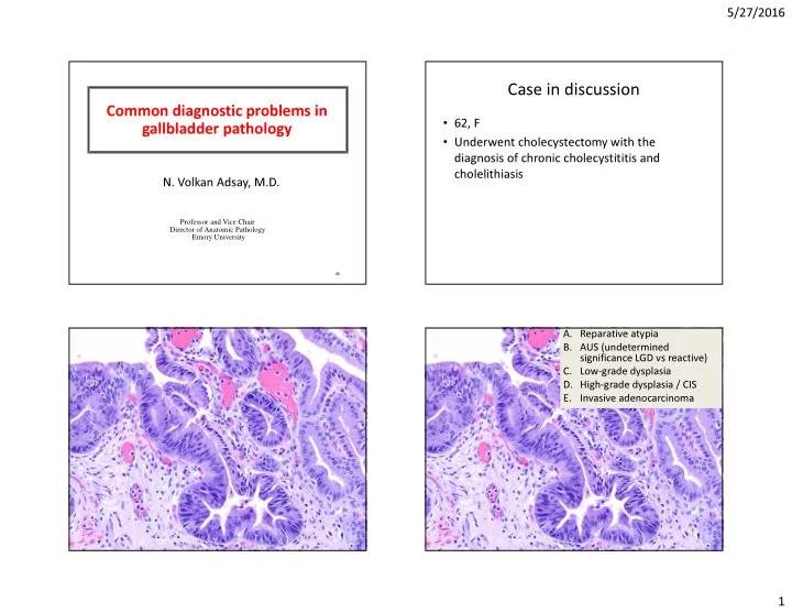
Case in discussion
- 62, F
- Underwent cholecystectomy with the
diagnosis of chronic cholecystititis and cholelithiasis
- A. Reparative atypia
- B. AUS (undetermined
significance LGD vs reactive)
- C. Low-grade dysplasia
- D. High-grade dysplasia / CIS
- E. Invasive adenocarcinoma