
24/01/2017 1
Bioburden-Biofilms in Inflammation
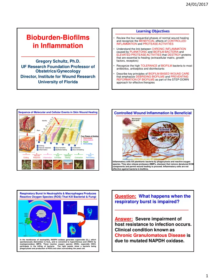
Gregory Schultz, Ph.D. UF Research Foundation Professor of Obstetrics/Gynecology Director, Institute for Wound Research University of Florida Learning Objectives
- Review the four sequential phases of normal wound healing
and recognize the BENEFICIAL effects of CONTROLLED INFLAMMATION and PROTEASE ACTIVITIES
- Understand the link between CHRONIC INFLAMMATION
caused by PLANKTONIC and BIOFILM BACTERIA and ELEVATED PROTEASE ACTIVITIES that DESTROY proteins that are essential to healing (extracellular matrix, growth factors, receptors)
- Recognize the high TOLERANCE of BIOFILM bacteria to most
antibiotics, antiseptics and disinfectants
- Describe key principles of BIOFILM-BASED WOUND CARE
that emphasize DEBRIDING BIOFILMS and PREVENTING REFORMATION OF BIOFILMS as part of the STEP-DOWN approach for effective therapies
1 2 3 4 5 6 7 . Vascular Scar Epithelial Scar Clotting Response Inflammation Formation Healing Contraction Remodeling
Sequence of Molecular and Cellular Events in Skin Wound Healing
Four Phases of Healing
- 1. Hemostasis
- 2. Inflammation
- 3. Repair
- 4. Remodeling
Inflammatory cells kill planktonic bacteria by phagocytosis and reactive oxygen
- species. They also release proteases (MMPs, elastase) that remove denatured ECM
components and permit wound healing to proceed. Inflammatory cells are not effective against bacteria in biofilms.
MMPs
Macrophages Neutrophils
ELASTASE
Controlled Wound Inflammation Is Beneficial
O2- H2O2 HOCl In the membranes of neutrophils, NADPH oxidase generates superoxide (O2-), which spontaneously dismutates to H2O2, and is converted to hypochlorous acid (HOCl) by myeloperoxidase (MPO). These reactive oxygen species (ROS), especially HOCl, participate in the killing of bacteria. The right panels show a bacteria being phagocytized and production of ROS (red color) surrounding the yeast cell.
Respiratory Burst In Neutrophils & Macrophages Produces Reactive Oxygen Species (ROS) That Kill Bacterial & Fungi