
SLIDE 1
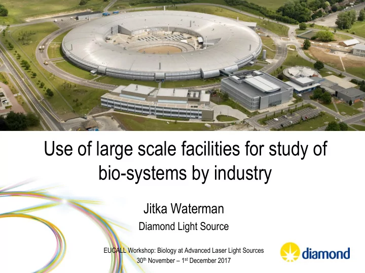
Use of large scale facilities for study of bio-systems by industry
Jitka Waterman
Diamond Light Source
EUCALL Workshop: Biology at Advanced Laser Light Sources 30th November – 1st December 2017
bio-systems by industry Jitka Waterman Diamond Light Source EUCALL - - PowerPoint PPT Presentation
Use of large scale facilities for study of bio-systems by industry Jitka Waterman Diamond Light Source EUCALL Workshop: Biology at Advanced Laser Light Sources 30 th November 1 st December 2017 Harwell Science & Innovation Campus MRC
EUCALL Workshop: Biology at Advanced Laser Light Sources 30th November – 1st December 2017
Diamond Light Source
ESA E6
RAL Space
ISIS - neutrons
Central Laser Facility
MRC PHE
Satellite Applications Catapult Research Complex
Phase 1 7 beamlines completed 2007 Phase 2 +15 beamlines completed 2012 Phase 3 +11 beamlines to be available by 2018
Key Facts Employee: > 500 External scientists: > 3000/year Publications: ~1000 in 2015 PDB: >4000 model deposited
Elizabeth Shotton Group Leader Anna Kroner XAS Leigh Connor XRPD, Engineering Claire Pizzey SAXS Alex Dias MX Jitka Waterman MX Renjie Zhang XChem Sin-Yuen Chang XAS Jason van Rooyen Cryo-EM Sally Irvine Imaging
Diamond – Opportunities related to structural biology
Macromolecular Crystallography Soft Condensed Matter Spectroscopy Materials Engineering and Environment Surfaces and Interfaces
I14
eBIC ePSIC
X-ray centring
sophisticated data collection with multi-axis goniometer functionality
profile, low resolution or large unit cell
– Project initiated in 2014 – Operational since 2015 – ILO commitment from early developments
– Rapid hit identification
Hits from JMJD2-DA (SGC)
Courtesy of Patrick Collins Diamond Light Source
Soaking compounds Crystal harvesting Data collection Hit identification
15-30 m 1-2 days 1-2 days 1/2 day
Crystal volume ~5000x smaller than 100 micron ’standard’ I sec exposure - 2Å data
– UK-XFEL Hub – Time resolved pump-probe experiments at slower time scale (ms) as an alternative to XFELs (fs) – LCP injector – sample preparation for XFEL experiment
‘Class C GPCR metabotropic glutamate receptor 5 transmembrane domain.’ Doré, AS et al. Nature (2014) 511, 557-562
by X-ray tomography
P12M detector Novel in-vacuum vessel
Tomography camera Goniometer Sample position Fluorescence detector OAV
Image-based alignment
FORMULATRIX
Automatic sample handling Compact hutch design
I13 I14 and EM Facility Building Services Diamond
Holbourn et al JBC (2011) 286 (25) 22243-22249
– Determine of the size and shape of proteins in solution – Low resolution 3D structure (~15 Å) – Map different components of a complex
– Ligand binding – Flexible proteins
– Investigate effect of different formulations – Protein stability studies
Parameters obtained by SAXS analysis
B23 experimental room
conformation under a range of conditions
the homogeneity of the sample preparation
structure content under high pressure
Rajasekaran et al, Biophys. Res. Com 398 (2010)
Protein stabilisation upon binding of a metal ion
Lipid Protein Nucleic acid & Carbohydrate Water
The mapping of an individual cell by SR-FTIR
– Imaging nanoparticles, for example determine location of particles in tumour cells – Could be used to image metal nanoparticles >20nm, might be challenging to view smaller/lighter particles
Nucleus
Nucleoli Nuclear membrane Mitochondria Nuclear pores ~ 120 nm in diameter
1 μm
EUCALL Workshop: Biology at Advanced Laser Light Sources 30th November – 1st December 2017