Basic Exam Skills Inspection Palpation Range of Motion Motor - - PowerPoint PPT Presentation

basic exam skills
SMART_READER_LITE
LIVE PREVIEW

Basic Exam Skills Inspection Palpation Range of Motion Motor - - PowerPoint PPT Presentation

12/12/2015 Basic Exam Skills Inspection Palpation Range of Motion Motor and Sensory Upper Extremity Exam: Vascular High Five!! Special tests Samantha Piper, MD Assistant Professor UCSF Hand and Upper Extremity


slide-1
SLIDE 1

12/12/2015 1

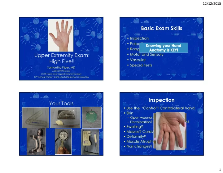
Upper Extremity Exam: High Five!!

Samantha Piper, MD

Assistant Professor UCSF Hand and Upper Extremity Surgery 10th Annual Primary Care Sports Medicine Conference

Basic Exam Skills

  • Inspection
  • Palpation
  • Range of Motion
  • Motor and Sensory
  • Vascular
  • Special tests

Your Tools Inspection

  • Use the “Control”! Contralateral hand
  • Skin

– Open wounds? Scars/marks? – Discoloration? Redness? Ecchymosis?

  • Swelling?
  • Masses? Cords?
  • Deformity?
  • Muscle Atrophy?
  • Nail changes?
slide-2
SLIDE 2

12/12/2015 2

  • Digital cascade/resting posture?

Inspection

Open wounds What lies beneath?

  • Location of wound + knowledge of

surface anatomy =

– Accurate assessment of potentially injured structures

  • Distal perfusion?

– Pulses, color, cap refill, temperature

  • Distal sensation and motor function?
  • Tendon continuity?

– Cascade, tenodesis, active movement

  • Bone or joint injury?

Inspection: Surface Landmarks

slide-3
SLIDE 3

12/12/2015 3

Palmar Landmarks

  • Name the

creases

Kaplan’s Cardinal Line

slide-4
SLIDE 4

12/12/2015 4

~1cm

~6mm

Flexor Surface Markers Dorsal Landmarks

slide-5
SLIDE 5

12/12/2015 5

Extensor Landmarks Palpation

  • Local Tenderness? – One finger
  • Abnormal bony anatomy?
  • Masses?
  • Joint effusion? Crepitus?
  • Pulses?
  • Temperature?

A1 Pulley: Trigger CMC: Carpal Boss MPJ: OA MP UCL

slide-6
SLIDE 6

12/12/2015 6

Thumb CMC Radial Styloid- Dequervain’s Scaphoid Lister’s Snuffbox

Scapho-styloid Joint Proximal Pole

Scaphoid- Tubercle

slide-7
SLIDE 7

12/12/2015 7

Lister’s SLJ Lunat e

Fovea PT Joint

DRUJ ECU LT Joint

Hook of Hamate

Range of Motion

  • Active & Passive
  • Is it limited? Why?

– Swelling? – Bone, Tendon, Nerve or Joint injury? – Arthritis? Disease?

  • Record the range of motion!

– Each Joint – Total Arc Motion (TAM) – Distance to Palmar Crease (DPC)

Extension Flexion Forearm P 70 S 85 Wrist 70, RD 20 75, UD 35 MP 0- +45 90 PIP 100 DIP 80 Thumb MP +10 55 Thumb IP +15 80 Thumb CMC PA 45 Thumb CMC RA 60

Testing Tendons

  • Tenodesis effect
slide-8
SLIDE 8

12/12/2015 8

FDS

  • Immobilize FDPs

FDP & FPL Terminal Tendon Central Slip

  • Elson Test

– PIP flexed 90* – Pt attempts to extend PIP – Assess DIP tension

  • Lax- CS intact
  • Tight- CS out
slide-9
SLIDE 9

12/12/2015 9

Strength

  • Test both hands, mean of 3 trials

– Grip

  • 5 adjustable hand spacings
  • Rapid alternating

– Pinch

  • Tip- M
  • Key- M&U

Upper Limb Neurologic Examination

  • Compression neuropathy- Always

check the C-spine!

  • Three major nerves

– Median – Ulnar – Radial

Cervical Radiculopathy

  • ROM in 3 planes (F/E, lateral F, Rot.)

– Pain or radicular radiation

  • TTP along C-spine
  • Spurling’s
  • Axial Compression

Two Point Discrimination

  • Innervation density
  • Objective
  • Test both sides of finger – different nerves
  • Normal < or = 5 mm

Thumb/Index/Long/Ring/Small 5/5/5/5/7 5r8u/7r5u/5/5/5

slide-10
SLIDE 10

12/12/2015 10

Median nerve - Motor

  • Extrinsic muscles: Origin in Forearm

– PT, FCR, PL – 4 FDS, 2 FDP (index & long) – FPL, PQ

  • Intrinsic muscles - Located in Hand

– Thenars (APB, FPB, Opp) – Lumbricals I, II

Median nerve – Extrinsic Motor

  • “OK” sign
  • AIN - Anterior Interosseous n.

OK! Not OK!

No DIP flexion!

Median nerve – Intrinsic Motor

  • Opposition

– “Make an L with your thumb”

Best Test

*

Palpate here -Feel the

Abductor Pollicis Brevis

Best Median Sensory

Palmar cutaneous nerve Common and Proper digital nerves

slide-11
SLIDE 11

12/12/2015 11

Ulnar nerve - Motor

  • Extrinsic – forearm origin

– FCU – 2 FDP (ring & small)

  • Intrinsic – located in the hand

– Hypothenars (ADM,FDM, ODM) – Lumbricals III & IV – Dorsal and palmar interosseous – Adductor Pollicis – Deep head FPB

Ulnar nerve – Extrinsic Motor

Small finger DIP flexion - FDP

Ulnar nerve – Intrinsic Motor

1st dorsal interosseous m. Hypothenar m.

Palpate here

Ulnar nerve- Intrinsic Motor

  • Wartenberg’s Sign

– Unopposed EDM (rad) due to loss of intrinsics

slide-12
SLIDE 12

12/12/2015 12

Ulnar intrinsic motor

  • Froment’s sign

– Substitute FPL for adductor

Ulnar nerve lesion

  • “Claw” deformity – “intrinsic minus”

Hyperextension of MP PIP Flexion

Best Ulnar Sensory

Common and Proper digital nerves

Radial nerve - Motor

  • All Extrinsic – from

forearm

  • Wrist, finger and

thumb extension

  • EPL: Retropulsion
  • PIP extension is

intrinsic function

– Median/ ulnar

slide-13
SLIDE 13

12/12/2015 13

Best Radial Sensory

Dorsal branches of proper digital nerves supply these areas

Digital Vascular Exam

  • Allen test
  • Doppler US of finger
  • O2 Sat Probe- should be NORMAL

Special Tests by Diagnosis Carpal Tunnel Syndrome

  • Tinel’s sign
  • Phalen’s test
  • Durkin’s carpal compression test
slide-14
SLIDE 14

12/12/2015 14

Tinel’s

  • Tap along course of nerve

– Positive if tingling in nerve distribution

Phalen’s test

Hold for 60s or until Sxs

Increases pressure on median nerve Positive if patient reports numbness in median distribution

Durkin’s Compression test

Hold for 30s or until Sxs

Positive if patient reports numbness in median distribution

Cubital Tunnel Syndrome

  • Tinel’s Sign
  • Flexion compression test
  • Ulnar nerve subluxation
slide-15
SLIDE 15

12/12/2015 15

Ulnar Nerve Flexion Compression Test

Hold for 30s or until sxs

Positive if patient reports numbness in median distribution

Thumb CMC Arthritis

  • Grind test – stresses joint

– Positive if painful

Thumb CMC Arthritis

  • Gelberman Adduction test –

stresses joint

– Positive if painful

Thumb CMC Arthritis

  • Shoulder sign
  • MP Hyperextension

– >30* is pathologic

slide-16
SLIDE 16

12/12/2015 16

Thumb & Finger MP CL tear

  • Varus and Valgus Stress test

– Thumb: 0 & 30* flexion – Digit: 0 & 90*

Positive if >30* angulation, no endpoint, or gross laxity compared to contralateral side

Wrist

Radial

  • Dequervain’s

– Finkelstein’s

  • SL Ligament

– Watson’s Shift

  • Ganglion Cyst

– Trans illumination

  • Scaphoid

injury/SLAC/SNAC Ulnar

  • DRUJ Instability

– Piano Key – Shuck

  • Ulnar Impaction/TFCC

– Ulnar carpal abutment

  • LT Ligament

– LT ballottement

  • ECU Pathology

– Subluxation – Synergy

DeQuervain’s tenosynovitis

Finkelstein’s test

Pain here!

SL Stability

  • Watson Shift test

Positive if asymmetrical click or if patient reports pain at DORSAL SLJ

slide-17
SLIDE 17

12/12/2015 17

Ganglion Cysts

  • Trans illumination
  • Don’t forget Allen’s if volar radial wrist!

DRUJ Instability

  • Piano Key

Positive if increased laxity compared to contralateral side

DRUJ Instability

  • Shuck

– Check in

  • Neutral
  • Supination
  • Pronation

Positive if increased laxity compared to contralateral side

Ulnar Impaction/TFCC

  • Ulnar carpal abutment stress test
slide-18
SLIDE 18

12/12/2015 18

ECU Pathology

  • ECU Synergy Test

– Resisted finger abduction – Positive if pain over ulnar wrist

ECU Pathology

  • ECU Stability

– Palpate ECU at ulnar groove through forearm ROM

  • Subluxates with Supination, Flexion, & UD
  • Reduces with pronation

– Positive if ECU felt subluxating out of groove, popping.

  • Resisted wrist extension and UD

– Pain indicated ECU pathology

Medial/Lateral Epicondylitis

  • Resisted wrist F/E with elbow

extended

– Positive if pain at M/L elbow

Thank You!

slide-19
SLIDE 19

12/12/2015 19

ELBOW Inspection: Carrying Angle

Normally:10* in men; 15* in women valgus deformity varus deformity

Inspection: Lateral Inspection

  • Posterior

– Olecranon bursa

  • Medial

– Subluxation of ulnar nerve

slide-20
SLIDE 20

12/12/2015 20

Inspection: Symmetry Bony Palpation Bony Palpation Bony Palpation

slide-21
SLIDE 21

12/12/2015 21

Bony Palpation Soft Tissue Palpation Range of Motion

Flexion >135 Extension 0 or more Compare to opposite side Pronation/supination ~80/~80 Compare to opposite side Keep elbows against flanks to block shoulder compensation

slide-22
SLIDE 22

12/12/2015 22

Functional Motion

  • Functional range E/F = 30-130*

– Loss of flexion more disabling than loss of extension

  • Functional range P/S = 50-50*

Neurologic Exam: Motor

  • Flexion: Brachialis, biceps (musc. n., C5,C6)
  • Extension: Triceps (radial n., C7)
  • Supination: Biceps, supinator (radial n., C6)
  • Pronation: Pronator teres (med. n., C6),

pronator quadratus (ain, C8, T1)

Neuro Exam:

Sensation

C5 C6 C7 C8 T1

Neuro Exam: Reflexes

  • Biceps - C5
  • Brachioradialis – C6
  • Triceps – C7
slide-23
SLIDE 23

12/12/2015 23

Special Tests

  • Tennis elbow

stress

‘Hook Test’ for distal biceps

O’Driscoll et al, AJSM 2007

Ligament Stability Lateral pivot shift test

(O'Driscoll/Morrey JBJS 73A:440,1991)

  • Supine
  • Extension
  • Supination
  • Valgus stress
  • Axial load

Apprehension or shift = ++ Subluxed in extension and reduces as you flex past 40-60 degrees

slide-24
SLIDE 24

12/12/2015 24

Pushup sign Chair sign

‘Moving Valgus Stress Test’ for MCL

  • Positive test is a subjective apprehension, instability, or

pain at the MCL origin between 70 & 120 degrees

  • 100% sensitive and 75% specific

O’Driscoll et al, AJSM 2007

Milking maneuver for MCL

  • Create a valgus stress by pulling on the patient's thumb with the

forearm supinated and elbow flexed at 90 degrees

  • Positive test is a subjective apprehension, instability, or pain at the

MCL origin

slide-25
SLIDE 25

12/12/2015 25 Thank You!