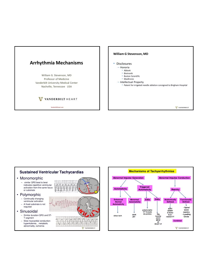

9/14/2019 William G Stevenson, MD Arrhythmia Mechanisms • Disclosures – Honoria • Abbott • Biotronik William G. Stevenson, MD • Boston Scientific • Medtronic Professor of Medicine – Intellectual Property Vanderbilt University Medical Center • Patent for irrigated needle ablation consigned to Brigham Hospital Nashville, Tennessee USA VanderbiltHeart.com Mechanisms of Tachyarrhythmias Sustained Ventricular Tachycardias • Monomorphic Abnormal Impulse Generation Abnormal Impulse Conduction – similar QRS beat to beat indicates repetitive ventricular Triggered activation from the same focus Automaticity Automaticity Reentry or substrate • Polymorphic – Continually changing Enhanced Abnormal DADs Anatomically Functionally EADs ventricular activation Normal Automaticity Defined Defined – A fixed substrate is not Automaticity required - Spiral AVRT • Sinusoidal wave polymorphic AVNRT VT Torsade reentry A flutter de pointes Dig - Leading – Similar duration QRS and ST- AIVR Scar- sinus tach toxicity JET related VT Circle T segment CPVT MFAT – Slow myocardial conduction: Combined AT hyperkalemia , metabolic RVOT VT abnormality, ischemia 1
9/14/2019 Early Afterdepolarizations Delayed Afterdepolarizations ▪ Occur after completion of repolarization ▪ Mechanisms: intracellular Ca+2 release leads to Yan et al Circulation 2001;103 increase Ca+2 efflux through the electrogenic ▪ Arise during plateau phase, prior to completion of Na+/CA2+ exchanger (NCX; 3Na : 1 Ca) causing a repolarization transient inward current ▪ Mechanisms: reactivation of ICaL or persistent ▪ Facilitated by cellular Ca+2 loading ▪ beta-adrenergic stimulation, rapid pacing activation of voltage gated Na channels during ▪ suppressed by adenosine through inhibition of prolonged AP ▪ Likely clinical manifestation: torsade de pointes, cAMP generation ▪ Arrhythmias: digoxin toxicity, CPVT, idiopathic polymorphic VT RVOT VT B Lerman Circ Arrhythm Ep 2015;8:483-491 A George JCI 2013 George JCI 2013 Maruyama et al Heart Rhythm 2014; Functionally defined reentry circuits Abnormal Automaticity leading circle reentry 0 mV abnormal normal automaticity ▪ Abnormal automaticity arises from partially Spiral Wave Reentry Reentry around a functional in cultured cardiomyocytes. depolarized cells area of block Rohr S Circ Arrhythm Electrophysiol . 2012;5:442 ▪ Sensitive to adrenergic stimulation, ▪ Does not require a structural substrate calcium channel blockers ▪ Reentry circuits can meander through the tissue or ▪ Not responsive to programmed stimulation or anchor on anatomic discontinuities (eg blood adenosine vessels) B Lerman. Circ Arrhythm Electrophysiol. 2015;8:483-491 2
9/14/2019 Polymorphic VT/VF with a structurally normal heart: Spiral Wave Reentry: - The sinus rhythm ECG may suggest a cause: - Functionally determined reentry - repolarization abnormalities - circuits can anchor on tissue discontinuity - possibly genetic - circuits can meander -waves can break up into daughter wavelets Short QT (genetic) In 3 dimensions a spiral wave is a scroll Initiation wave Long QT Early (genetic ) block repolarization Brugada (genetic) AE Panfilov et al Spiral Wave Reentry www- in cultured cardiomyocytes. KLEBER, A. G. et al. Physiol. Rev. binf.bio.uu.nl/~panfil 84: 431-488 2004 Catecholaminergic polymorphic VT Rohr S Circ Arrhythm ov/research.html Electrophysiol . 2012;5:442 (genetic, normal resting ECG) Scar-related Reentrant VT Initiation of scar- related reentry monomorphic VT sinus rhythm • VT substrate is “ stable ” Focal region of • Repeated VT episodes may occur over yrs conduction block Slow conduction • VT is inducible at EP study to allow • Drug efficacy for prevention is poor recovery from block 3
9/14/2019 Slow conduction: Zig - zag conduction caused by fibrotic As cell to cell coupling decreases, conduction slows but separation of myocyte bundles the “safety factor” for conduction increases because De Bakker, et al. Circulation 1988; 77:589. Circulation 1993;88:915. less current is dissipated to surrounding cells sink source - - - - - - - + + + + + + + Safety factor – excess current Fibrosis Myocyte + + + + + + + - - - - - - - available above that required bundles for conduction intracellular current less likely to block Sa well coupled poorly coupled Human papillary muscle in cross section Unidirectional conduction block due Fibrosis in ventricular scars: Anatomy to source sink mismatch defines reentry paths, slows conduction, facilitates small source large source block Geometry of large sink small sink Myocytes in infarct fibrosis myocytes surrounded by Fibroblasts / myofibroblast as fibrosis bridges that slow conduction? myocytes fibroblasts myocytes promotes conduction block Stable conduction Time (ms) variations in geometry can promote Dense fibrosis unidirection conduction block and reentry Gaudesius et al. Circ Res 2003;93:421 Magoli et al Circ Res 2006; 98:801 Wijnmaalen A P et al. Nguyen, Qu, Weiss. Fibrosis and Circulation Smaill B H et al. Circulation Roden and Tomaselli. Conceptual basis for arrhythmology 2005 arrhythmogensis. J Mol Cell Cardiol 2014 Research . 2013;112:834-848 2010;121:1887 4
9/14/2019 Sustained monomorphic VT in non-ischaemic Remodeling of Cardiac Scars is Dynamic cardiomyopathy is associated with fibrosis Glashan and Zeppenfeld et al EHJ 2018 Fibroblast proliferation Myocyte Myocyte Hypertrophy Death Myofibroblasts Matrix Synthesis of extracellular matrix metaloproteinases proteins replacement fibrosis degradation of ECM proteins variable patterns of fibrosis Fibrosis: interstitial / replacement - impaired contraction - impaired relaxation - cellular uncoupling - reduced capillary density - Electrophysiologic effects: conduction slowing and block Burchfield et al Circulation 2013; 128:388. Brown. Cardiac extracellular matrix: a dynamic entity. AJP Heart Circ Physiol 289: 2005; Nonischemic Cardiomyopathy Sustained Monomorphic Reentry circuits defined by functional VT: Scar location predicts VT morphologies, ablation block and wave collision approach and outcomes Basal Septal - Antero Septal Scar Basal Lateral LV Scar A stable reentry circuit isthmus can be defined by slow propagation Exit high transverse to fiber curvature orientation and wavefront radius collision. Slow conduction occurs at areas of high wavefront curvature at entrance and exit regions Entrance Septal ablation is required Basal lateral LV ablation required - Epicardial ablation not usually helpful - Epicardial ablation often needed - Risk of AV block - Lower efficacy? High resolution mapping and entrainment of reentry Anter et al Circulation 2016 Piers S R et al. Circ Arrhythm Electrophysiol . 2013. Ororiz et al Circ Arrhythm Electrophysiol 2014; in 6 – 8 wk old swine infarct model 5
9/14/2019 Propagation during VT Voltage map Fibrosis: Diminished Myocyte Coupling • Slow conduction • Unidirectional conduction block • Promotes automaticity – makes it easier for automaticity to overcome electronic effects of neighboring cells Myocardial Fibrosis: a substrate for ventricular arrhythmias • Myocardial infarction • Dilated cardiomyopathies Thank You • ARVC/D • Inflammatory cardiomyopathies – Sarcoidosis, Chagas, post myocarditis • Surgically repaired congenital heart disease • Mitral valve prolapse • Elderly, hypertension • Elite athletes • LV assist devices 6
Recommend
More recommend