Antigen Effector Antigen uptake function presentation Dendritic - - PowerPoint PPT Presentation

antigen effector antigen uptake function presentation
SMART_READER_LITE
LIVE PREVIEW

Antigen Effector Antigen uptake function presentation Dendritic - - PowerPoint PPT Presentation

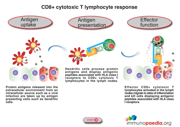
CD8+ cytotoxic T lymphocyte response Antigen Effector Antigen uptake function presentation Dendritic cells process protein antigens and display antigenic peptides associated with HLA class I receptors to CD8+ cytotoxic T lymphocytes in


slide-1
SLIDE 1

CD8+ cytotoxic T lymphocyte response

START

Protein antigens released into the extracellular environment from an intracellular source such as a viral infection are taken up by antigen presenting cells such as dendritic cells. Dendritic cells process protein antigens and display antigenic peptides associated with HLA class I receptors to CD8+ cytotoxic T lymphocytes in the lymph nodes. Effector CD8+ cytotoxic T lymphocytes activated in the lymph nodes migrate to sites of inflammation and kill cells displaying antigenic peptides associated with HLA class I receptors.

Antigen presentation Antigen uptake Effector function

slide-2
SLIDE 2

Phagocytosis by dendritic cells

Dendritic cells are specialised antigen presenting cells derived from monocytes which develop in the bone marrow from long- lived haematopoietic stem cells committed to a myeloid lineage. Dendritic cells engulf foreign antigens and micro-organisms in the extracellular environment and process peptide for antigen presentation. The activated dendritic cells migrate to the T cell zone of secondary lymphoid organs where they present peptide antigens associated with cell surface HLA class I and II receptors to T lymphocytes. Tissue cells Bone marrow Monocyte Dendritic cell Secondary lymphoid

  • rgan

T cell zone

Phagocytosis

Virus

PREVIOUS NEXT Antigen uptake

slide-3
SLIDE 3

Generation of naive T lymphocytes

T lymphocytes develop in the bone marrow from long-lived haematopoietic stem cells committed to a lymphoid lineage. They migrate to the thymus where through gene rearrangements each T cell expresses a unique T cell receptor (TCR) destined to recognise a single peptide epitope. T cells also express either the CD4 or CD8 surface molecules. Non-functional and “self”- reactive T cells are deleted in the thymus by positive and negative selection processes. Naive T lymphocytes leave the thymus and circulate to the T cell zone of secondary lymphoid organs where they can be stimulated by dendritic cells presenting peptide antigens associated with cell surface HLA class I and II receptors. Naive CD8+ cytotoxic T lymphocyte Naive CD4+ helper T lymphocyte TCR CD4 TCR CD8 Bone marrow Thymus Secondary lymphoid

  • rgan

T cell zone

PREVIOUS NEXT Antigen presentation

slide-4
SLIDE 4

Priming of CD8+ cytotoxic T lymphocytes

Activated dendritic cells present peptide antigens associated with cell surface HLA class I receptors to CD8+ cytotoxic T lymphocytes in the T cell zone of secondary lymphoid organs. They are able to prime naive CD8+ cytotoxic T lymphocytes expressing cell surface T cell receptors (TCR) that recognise the peptide antigens displayed. Once primed, CD8+ cytotoxic T lymphocytes require activation signals from activated CD4+ helper T lymphocytes are in order to develop into killing cells. Secondary lymphoid

  • rgan

T cell zone Naive CD8+ cytotoxic T lymphocyte Activated dendritic cell

PREVIOUS NEXT

TCR HLA I CD8 T cell priming signal

Antigen presentation

slide-5
SLIDE 5

Activation of CD8+ cytotoxic T lymphocytes

Primed CD8+ cytotoxic T lymphocytes acquire activation signals from activated CD4+ helper T lymphocytes. Following activation, CD8+ cytotoxic T lymphocytes proliferate and differentiate into effector cells capable of killing infected cells. Effector cells migrate to sites of inflammation. Memory CD8+ cytotoxic T lymphocytes cells also develop to establish long-term immunity. Secondary lymphoid

  • rgan

T cell zone Primed CD8+ cytotoxic T lymphocyte Activated CD4+ helper T lymphocyte CD4 TCR T cell activation signal

PREVIOUS NEXT

TCR CD8

Antigen presentation

slide-6
SLIDE 6

HLA class I antigen presentation by infected cells

Cells infected by viruses or intracellular bacteria and also certain types of tumour cells are detected and destroyed by CD8+ cytotoxic T lymphocytes. CD8+ cytotoxic T lymphocytes are activated in the secondary lymphoid organs and migrate to sites

  • f inflammation where they scan cell surfaces with their T cell receptors (TCR) for recognition of foreign peptide antigens

displayed on cell surface HLA class I receptors. HLA class I receptors are constitutively expressed on the surface of all cells except erythrocytes. CD8+ cytotoxic T lymphocytes destroy target cells by releasing perforin and cell degrading molecules.

HLA I TCR

CD8

Kill

How are HLA class I antigens processed? Click here. Tissue cells Viral protein

Virus

“Self” protein Activated CD8+ cytotoxic T lymphocyte

Effector function PREVIOUS NEXT

slide-7
SLIDE 7

HLA class I antigen presentation by healthy cells

Effector CD8+ cytotoxic T lymphocytes that are activated in the secondary lymphoid organs migrate to sites of inflammation where they scan cell surfaces with their T cell receptors for recognition of foreign peptide antigens associated with HLA class I receptors. HLA class I receptors are constitutively expressed on the surface all cells except erythrocytes. “Self”-peptides are not normally recognised due to prior deletion of auto-reactive T lymphocytes in the thymus. Tissue cells Activated CD8+ cytotoxic T lymphocyte HLA I “Self” peptide “Self” protein TCR CD8

No Recognition

How are HLA class I antigens processed? Click here.

Effector function PREVIOUS BACK TO START