Antigen Antigen Effector uptake presentation function Protein - - PowerPoint PPT Presentation

antigen antigen effector uptake presentation function
SMART_READER_LITE
LIVE PREVIEW

Antigen Antigen Effector uptake presentation function Protein - - PowerPoint PPT Presentation

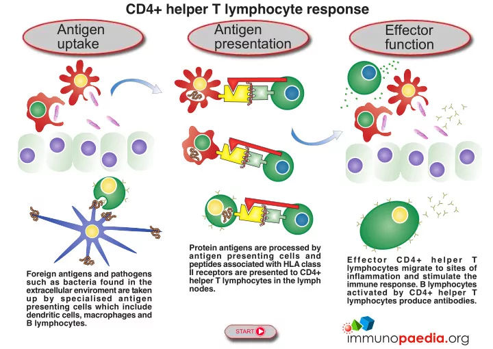
CD4+ helper T lymphocyte response Antigen Antigen Effector uptake presentation function Protein antigens are processed by antigen presenting cells and E f f e c t o r C D 4 + h e l p e r T peptides associated with HLA class lymphocytes


slide-1
SLIDE 1

START

CD4+ helper T lymphocyte response

Foreign antigens and pathogens such as bacteria found in the extracellular enviroment are taken up by specialised antigen presenting cells which include dendritic cells, macrophages and B lymphocytes. Protein antigens are processed by antigen presenting cells and peptides associated with HLA class II receptors are presented to CD4+ helper T lymphocytes in the lymph nodes. E f f e c t o r C D 4 + h e l p e r T lymphocytes migrate to sites of inflammation and stimulate the immune response. B lymphocytes activated by CD4+ helper T lymphocytes produce antibodies.

Antigen presentation Antigen uptake Effector function

slide-2
SLIDE 2

Phagocytosis by macrophages and dendritic cells

Dendritic cells and macrophages are specialised antigen presenting cells derived from monocytes which develop in the bone marrow from long-lived haematopoietic stem cells committed to a myeloid lineage. Dendritic cells and macrophages engulf foreign proteins and micro-organisms in the extracellular environment and process peptide antigens. The activated dendritic cells and macrophages migrate to the T cell zone of secondary lymphoid organs where they present peptide antigens associated with cell surface HLA class II receptors to CD4+ helper T lymphocytes. Tissue cells Bone marrow Monocyte Dendritic cell Secondary lymphoid

  • rgan

T cell zone Phagocytosis Bacterium Macrophage

PREVIOUS NEXT Antigen uptake

slide-3
SLIDE 3

Endocytosis by B lymphocytes

B lymphocytes produce antibodies and are also antigen presenting cells. They develop in the bone marrow from long-lived haematopoietic stem cells committed to a lymphoid lineage. They migrate from he bone marrow to the B cell zone (germinal centre) of secondary lymphoid organs where they contact follicular dendritic cells carrying antigens on their cell surface. Recognition of antigen by the B cell receptor (BCR) initiates endocytosis followed by peptide processing. The B cell is now primed for antibody production and migrates from the B cell zone to the T cell zone for presentation of peptide antigens associated with cell surface HLA class II receptors to CD4+ helper T lymphocytes. Bone marrow Secondary lymphoid

  • rgan

B cell zone

PREVIOUS NEXT

Naive B lymphocyte Secondary lymphoid

  • rgan

T cell zone Follicular dendritic cell Protein antigen Primed B lymphocyte BCR

Antigen uptake

Endocytosis

slide-4
SLIDE 4

Generation of naive CD4+ helper T lymphocytes

Naive CD4+ helper T lymphocyte CD4

TCR

Bone marrow Thymus Secondary lymphoid

  • rgan

T cell zone CD4+ helper T lymphocytes develop in the bone marrow from long-lived haematopoietic stem cells committed to a lymphoid

  • lineage. They migrate to the thymus where through gene rearrangements each T cell expresses a unique T cell receptor (TCR)

destined to recognise a single peptide epitope. The T cells also express the CD4 surface molecule. Non-functional and “self”- reactive T cells are deleted by processes of positive and negative selection. Naive CD4+ helper T lymphocytes leave the thymus and circulate to the T cell zone of secondary lymphoid organs for stimulation by dendritic cells presenting peptide antigens associated with cell surface HLA class II receptors.

PREVIOUS NEXT Antigen presentation

slide-5
SLIDE 5

Activation of CD4+ helper T lymphocytes by dendritic cells

CD4+ helper T lymphocytes are activated by dendritic cells presenting peptide antigens associated with HLA class II receptors in the T cell zone of secondary lymphoid organs. Activated CD4+ helper T lymphocytes proliferate and migrate to sites of

  • inlammation. Memory CD4+ helper T lymphocytes are also generated for long-term immunity.

T cell zone Secondary lymphoid

  • rgan

T cell activation signal How are HLA class II antigens processed? Click here. CD4+ helper T lymphocyte CD4

HLA II

Activated dendritic cell

TCR

PREVIOUS NEXT Antigen presentation

slide-6
SLIDE 6

Activation of CD4+ helper T lymphocytes by macrophages

CD4+ helper T lymphocytes are activated by macrophages presenting peptide antigens associated with HLA class II receptors in the T cell zone of secondary lymphoid organs. T cell zone Secondary lymphoid

  • rgan

T cell activation signal Activated macrophage CD4

TCR HLA II

CD4+ helper T lymphocyte How are HLA class II antigens processed? Click here.

PREVIOUS NEXT Antigen presentation

slide-7
SLIDE 7

Activation of B lymphocytes by CD4+ helper T lymphocytes

CD4+ helper T lymphocytes stimulate B lymphocytes presenting peptide antigens associated with HLA class II receptors in the T cell zone of secondary lymphoid organs. The CD4+ helper T lymphocyte provides activation signals to the B lymphocyte to proliferate and differentiate into antibody-secreting cells (plasma cells). Memory B lymphocytes are also generated for long- term immunity. T cell zone Secondary lymphoid

  • rgan

Antibodies

PREVIOUS NEXT

B cell activation signal Activated B lymphocyte

TCR HLA II

CD4+ helper T lymphocyte How are HLA class II antigens processed? Click here. Plasma cell

CD4

Antigen presentation

slide-8
SLIDE 8

Immune stimulation by CD4+ helper T lymphocytes

Activated CD4+ helper T lymphocytes migrate from the secondary lymphoid organs to sites of inflammation where they stimulate the immune reponse by release of inflammatory cytokines. Tissue cells Dendritic cell Phagocytosis Bacterium Macrophage

PREVIOUS

Activated CD4+ helper T lymphocyte Antibodies

Effector function BACK TO START