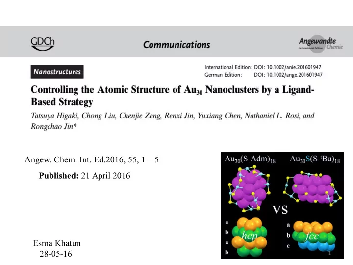
Angew. Chem. Int. Ed.2016, 55, 1 5 Published: 21 April 2016 Esma - - PowerPoint PPT Presentation

angew chem int ed 2016 55 1 5 published 21 april 2016
SMART_READER_LITE
LIVE PREVIEW

Angew. Chem. Int. Ed.2016, 55, 1 5 Published: 21 April 2016 Esma - - PowerPoint PPT Presentation

Angew. Chem. Int. Ed.2016, 55, 1 5 Published: 21 April 2016 Esma Khatun 28-05-16 1 Introduction Atomically precise nanoclusters (NCs) are missing link between small molecules and nanoparticles. Investigation of size dependent


slide-1
SLIDE 1

Published: 21 April 2016

1

  • Angew. Chem. Int. Ed.2016, 55, 1 – 5

Esma Khatun 28-05-16

slide-2
SLIDE 2

In this paper …

 They have reported X-ray single crystal structure of Au30(S-Adm)18 and compared this structure with previously reported Au30S(S-tBu)18.  They have provided a method for the discovery of possibly overlooked clusters because of their anomalous solubility.

Introduction

  • Atomically precise nanoclusters (NCs) are missing link between small molecules and

nanoparticles.

  • Investigation of size dependent properties of NCs is important step towards understanding

their potential applications such as catalysis.

  • The relationship between the structure of NCs and the protecting ligand still remains elusive.
  • Ligand bulkiness influence the structure and sizes of NCs, whereas the aromaticity of

ligands seems less critical.

  • It was also found that the substituted benzenethiolates provide an effective means for

tailoring the size and structure of NCs.

2

slide-3
SLIDE 3

3

UV-Vis spectra of (A) Au30(S-Adm)18 and (B) Au30(StBu)18 and Au30S(S-tBu)18

HAuCl4·3 H2O + [(C8H17)4N]+Br methanol

HS-Adm

Further reduction with NaBH4 Stirred over a period of one week Washed the crude product with methanol and dichloromethane three times Pure Au30(S-Adm)18 NCs were extracted with benzene Single-crystal growth was performed by vapor diffusion of cyclohexane into a benzene solution of the NCs

slide-4
SLIDE 4

4

Time course of UV-Vis spectra of crude Au30(S- Adm)18 NC. Time course of MALDI-MS spectra of crude Au30(S-Adm)18 NC.

slide-5
SLIDE 5

5

The effect of DCM washing monitored with MALDI-MS MALDI mass spectrum of pure Au30(S-Adm)18.

slide-6
SLIDE 6

6

Overall structure of the Au30(S-Adm)18 NC : A) Unit cell with a FCC superlattice arrangement ; B) top view; C) side view. Labels: magenta=Au, yellow=S, gray=C, white=H. The carbon tails are in wireframe mode

slide-7
SLIDE 7

7

Anatomy of the structure of Au30(S-Adm)18 NC: A,B) Top view of the Au18 kernel and the addition

  • f six dimeric staple motifs in two steps (in blue and green, respectively); C,D) Side view of the

kernel and addition of six staples; E) Four-layer structure of the Au18 kernel in a HCP manner; F,G) Six Au4 assembled pattern in top and side views. Color labels: magenta=Au in the kernel, light blue=Au in the staple, yellow=S.

slide-8
SLIDE 8

Au-Au bond length distribution in the Au30(S-Adm)18 nanocluster

8

slide-9
SLIDE 9

9

Comparison of the Au30(S-Adm)18 and Au30S(S-tBu)18 structures (carbon tails omitted). A) Au18 kernel and staples; B) Au18 core only; C) Au18 core of HCP arrangement in Au30(S-Adm)18; and D) Au22 core and staples; E) Au22 core only; F) Au22 core of FCC arrangement in Au30S(S-tBu)18

slide-10
SLIDE 10

10

Conclusion

  • They reported first example of gold NCs packed into an FCC superstructure.
  • Structural control of the Au30 NC is realized by exploring the bulky adamantanethiol

ligand.

  • The new cluster was obtained in 20 % yield (Au atom basis) via spontaneous size

focusing under mild conditions that do not lead to any sulfido ligand as in the previously reported Au30S(StBu)18 and Au38S2(S-Adm)20.

  • The newly obtained Au30(S-Adm)18 nanocluster shows distinct optical absorption

features and peculiar solubility.

  • The structure of Au30(S-Adm)18 is rather different from the previously reported

Au30S(StBu)18 cluster and also the theoretical structures of Au30(SH)18 and Au30(StBu)18.

slide-11
SLIDE 11

11

DOI:10.1021/acs.chemmater.5b05008

  • Chem. Mater. March 24, 2016, 28, 3292−3297
slide-12
SLIDE 12

12

Introduction

  • The typical routes for metal NC synthesis are (i) direct reduction of metal precursors in the

presence of desired ligands, (ii) postsynthetic ligand-exchange (LE), and (iii) metal- exchange.

  • LE has recently been widely adopted for preparing novel gold NCs, but the use of LE in

silver is rare, and its mechanism remains unexplored and poorly understood.

  • Particularly in silver, LE is exclusively based on a biphasic approach, where the starting and

the final products move between two immiscible phases.

  • In this paper they designed a single phase LE reaction to understand the step-by-step

conversion of Ag44(SR)30 NCs to Ag25(SR)18 NCs, and vice versa.

  • The single phase LE of Ag44(SPhF)30 clusters (SPhF: 4-fluorobenzenethiolate) with 2,4

dimethylbenzenethiol (HSPhMe2) initially led to the partial LE and then complete LE. After complete LE, the Ag44 experienced structural distortions to form Ag25(SPhMe2)18 and intermediate NCs of sizes larger than Ag44.

  • The formation of Ag44(SPhF)30 from the Ag25(SPhMe2)18 was observed to occur by

dimerization of Ag25 followed by rearrangements.

slide-13
SLIDE 13

UV−vis of Ag44(SPhCOOH)30 and its HSPhMe2- exchanged product (red trace).

Negative mode ESI MS of (A) Ag44(SPhCOOH)30 and (B) its ligand- exchange (LE) product with HSPhMe2. The assigned peak of [Ag44(SPhCOOH)30]4- is due to as synthesized product and the other [Ag44(SPhCOOH)29]4- is due to a fragment of parent cluster.

UV-vis absorption spectra of Ag25(SPhMe2)18 and its HSPhCOOH exchanged product Ag44(SPhCOOH)30

13

slide-14
SLIDE 14

Time-dependent absorption study of Ag44(SPhF)30 LE with HSPhMe2 Time-dependent ESI MS study of [Ag44(SPhF)30]4– LE with HSPhMe2. The numbers with black, red, and blue color correspond to the number of Ag atoms, SPhF, and SPhMe2 ligands,

  • respectively. The numbers in green represent the charge of the clusters. Sharp peaks labelled

with “$” are unknown impurity artifacts in the mass spectrometer without silver isotopic pattern, which are also present in the spectrum of a pure HPLC-grade DCM control. Mass spectral data reveal the conversion of Ag44 to Ag25 through intermediate NCs.

14

slide-15
SLIDE 15

15

(A) Time-dependent ESI MS. Sharp peaks labeled with“$” are unknown impurity artifacts in the mass spectrometer without silver isotopic pattern, which are also present in the spectrum of a pure HPLC-grade DCM control (B) UV−vis study of Ag25(SPhMe2)18 LE with

  • HSPhF. Red asterisk in B (on blue curve)

shows the appearance of Ag44 features. Inset: photographs of Ag25(SPhMe2)18 LE as a function of time. Color change from orange Ag25 to red indicates the formation of Ag44 cluster

slide-16
SLIDE 16

16

All color spheres: Ag atoms. Left half cycle: stage 1, partial LE; stage2, nearly complete LE; stage3, disproportionation; and stage 4, size focusing. Right half cycle: stage 1, partial LE; stage 2, dimerization; stage 3, disproportionation; and stage 4, size focusing

slide-17
SLIDE 17

17

Conclusion

  • They have successfully designed a single phase LE reaction to completely elucidate

the step-by-step transformation of Ag44(SR)30 NCs to Ag25(SR)18 NCs, and vice versa.

  • They have done detailed investigations of the LE with unprecedented atomic detail

reveal that the Ag44 to Ag25 transformation occurs via a disproportionation mechanism, whereas its reverse occurs through an uncommon dimerization prior to disproportionation.

slide-18
SLIDE 18

18

Future plan

Ag54 2 , 4

  • H

S P h M e2 Ag25 1,3-BDT Ag29 2 , 4

  • H

S P h C l 2 , 4

  • F

T P Ag44

  • As adamentanethiol produce some

unusual structure, so we can try ligand induced reaction of Ag54 with this

  • thiol. It may form new silver cluster
  • f interesting structural properties.
slide-19
SLIDE 19

19

Bader charge analysis on the total structure of Au30S(S-tBu)18 (A) and the DFT-derived structure

  • f Au30(S-t-Bu)18 (B). The“19th sulfur ” is shown by the arrow in (A). Colors of atoms, t-Bu blue

sticks, sulfur yellow, gold in ranging colors from blue (negative) to red (positive)