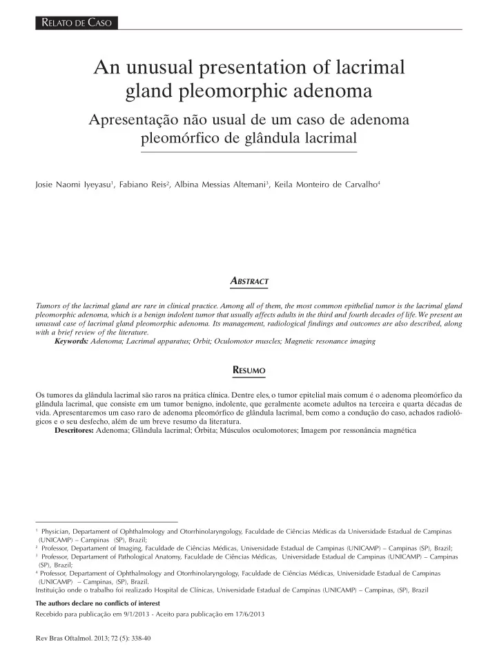
SLIDE 3 340 Rev Bras Oftalmol. 2013; 72 (5): 338-40 Figure 5: Areas with myxoid stroma and typical cellular components (ductal structures and myoepithelial cells) Iyeyasu JN, Reis F , Altemani AM, Carvalho KM
images and heterogeneously hyperintense on T2-weighted images, due to higher water content, with moderate contrast enhancement after gadolinium-DTPA injection(9). Although not usual, there may be intratumoral hemorrhage(7), bone destruction(4,9) and calcification(4,9,10). Intracranial extension, as happened in our case, is very rare. Although the tumor presents characteristic clinical and radiological features, which usually allows preoperative diagnosis(4,5,11), the definitive diagnosis is based on the histopathological examination(11), which shows cords of well- differentiated epithelial tubules derived from the ducts of the lacrimal gland, within loose myxomatous connective tissue(11). In
- ur case, due to the clinical features and the extension of the
tumor, a malignant tumor (epithelial carcinoma) would be the most likely diagnosis, however, an extensive histopathological examination was performed and no features of malignancy was found, confirming the diagnosis of LGPA. Differential diagnosis includes lymphoma, chronic dacryoadenitis, Sjogren’s syndrome, adenoid cystic carcinoma, granulomatous dacryoadenitis (sarcoidosis), benign lymphoid hyperplasia, cavernous hemangioma(5), intralacrimal schwannoma and hemangiopericytoma(6). The treatment is the complete excision of the tumor and adjacent tissues, usually by lateral orbitotomy. It is believed that preoperative biopsy and incomplete resection could lead to the tumor recurrence (even after years), as well as to malignant transformation(2-7). Complications following surgery include orbital hemorrhage, edema, optic nerve compression, orbital infection, lateral gaze palsy(2), dry eye, ptosis, lid retraction and transient diplopia(5). The prognosis is good when the lesion is completely excised with an intact capsule(3,4). A recurrence rate of 3% within five years has been reported in complete excisions and a recurrence rate of 32% over 15 years in incomplete ones(2,3,7). It is estimated that 10% of pleomorphic adenomas undergo malignant change within 20 years after first treatment and 20% by the end of 30
- years. Malignant transformation of a benign pleomorphic
adenoma into a squamous cell carcinoma has also been reported 19 years after the initial operation(3). We consider our case unusual due to its presentation at an
- lder age, with bone destruction, invasion of the extraocular
muscles and intracranial extension, which are rare features in cases of LGPA. In other words: it seems to be a malignant tumor, but it is not.
REFERENCES
1. Santos RR, Damasceno RW, de Pontes FS, Cursino SR, Nishiwaki- Dantas MC, Vital Filho J, et al. Ten-year follow-up of a case series of primary epithelial neoplasms of the lacrimal gland: clinical features, surgical treatment and histopathological findings. Arq Bras Oftalmol. 2010;73(1):33-9. 2. Halli RC, Mishra S, Kini YK, Kharkar VR, Hebbale MA. Modified lateral orbitotomy approach: a novel technique in the management of lacrimal gland tumors. J Craniofac Surg. 2011;22(3): 1035-8. 3. Chandrasekhar J, Farr DR, Whear NM. Pleomorphic adenoma of the lacrimal gland: case report. Br J Oral Maxillofac Surg. 2001;39(5):390-3. 4. Friedhofer H, Mendonça FP, Salles AG, Ferreira MC. Adenoma pleomórfico de glândula lacrimal - Relato de caso. Rev Soc Bras Cir
5. Prabhakaran VC, Cannon PS, McNab A, Davis G, O’Donnell B, Dol- man PJ, et al. Lesions mimicking lacrimal gland pleomorphic adenoma. Br J Ophthalmol. 2010;94(11):1509-12. 6. Rose GE. To crash or not to crash? Probability in the management of benign lacrimal gland tumours. Eye (Lond). 2009;23(8): 1625-8. 7. Miyazaki T, Yamasaki T, Moritake K, Matsumoto Y, Akiyama Y, Nagai H, et al. Unusual progression of pleomorphic adenoma of the lacrimal gland: case report. Neurol Med Chir (Tokyo). 2005;45(8):407-10. 8. Gupta S, Garg S, Singh S, Hasija S, Chaudhary M. Pleomorphic ad- enoma of lacrimal gland in a 5-year-old child: Diagnosed on aspira- tion cytology. Diagn Cytopathol. 2013;41(6):565-6. 9. Gündüz K, Shields CL, Günalp I, Shields JA. Magnetic resonance im- aging of unilateral lacrimal gland lesions. Graefes Arch Clin Exp
- Ophthalmol. 2003;241(11):907-13.
10. McNab AA, Satchi K. Recurrent lacrimal gland pleomorphic ad- enoma: clinical and computed tomography features. Ophthalmology. 2011;118(10):2088-92. 11. Ostrosky A, Klurfan FJ, Gonzalez MJ, Camaly D, Villa D. Pleomor- phic adenoma of the lacrimal gland. Case report. Med Oral Patol Oral Cir Bucal 2005;10(1):88-9; 86-8.
Author corresponding Fabiano Reis Hospital de Clínicas da Universidade Estadual de Campinas (HC - Unicamp) Rua Vital Brasil, nº 251 – Cidade Universitária Zeferino Vaz Zip code 13083-888 – Campinas (SP) – Brazil Postal code 6142 Phone: 55-19-35217280 E-mail: fabianoreis2@gmail.com