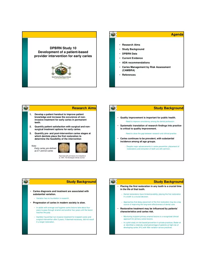

Agenda • Research Aims DPBRN Study 10 • Study Background Development of a patient-based • DPBRN Data provider intervention for early caries • Current Evidence • ADA recommendations • Caries Management by Risk Assessment (CAMBRA) • References Research Aims Study Background 1. Develop a patient handout to improve patient knowledge and increase the occurence of non- • Quality improvement is important for public health. invasive treatment for early caries in permanent - Need to improve consistency among the dental profession teeth. • Systematic translation of research findings into practice 2. Quantify patient satisfaction with surgical and non- is critical to quality improvement. surgical treatment options for early caries. 3. Quantify pre- and post-intervention caries stages at - Need to close the gap between research and clinical practice which dentists place the first restoration to • Caries continues to be prevalent, with substantial determine the feasibility of the intervention incidence among all age groups. Note: - Despite major advancements in caries prevention, placement of Early caries are defined restorations and extraction of teeth are still common. as E1 and E2 caries Reprinted with permission from Espelid et al, 1997, The Norwegian Dental Journal. Study Background Study Background Placing the first restoration in any tooth is a crucial time • in the life of that tooth. • Caries diagnosis and treatment are associated with substantial variation. – Dental restorations have limited durability; placing the first restoration in a tooth is a crucial decision. – Variation has no foundation in research. – Approaches that delay placement of the first restoration may be a key • Progression of caries in modern society is slow. source of improving the long-term effectiveness of dental care . – In adults with average oral hygiene caries lesions take about four • Restorative treatment may be influenced by patients’ years to pass through enamel and another four years until the lesion characteristics and caries risk. reaches the pulp. – Monitoring incipient primary enamel lesions is a recognized clinical – Hamilton found that non-invasive treatment for incipient caries and approach for primary caries lesions. surgical intervention after 2 years, if deemed necessary, did not result in a larger restoration. – In a pilot study on risk-based prevention in private practices, Bader et al. identified a relatively small percentage of patients at high-risk of developing caries (4%) with little variation across practices.
DPBRN Data Study Background • Patient satisfaction is important because it leads to DPBRN Study: Reasons for Placing Restorations on quality improvement. Previously Unrestored Permanent Tooth Surfaces – Medical professionals’ perceptions and patients’ perceptions about treatment they receive differ. • Objectives of interest for the current study: – Patient satisfaction is linked to regular return visits, caregiver trust, – To identify the reasons that dentists place perception of technical competence, and treatment outcomes. restorations in unrestored tooth surfaces. • Patient education and decision aids can improve the provider-patient relationship, decision-related – To assess pre- and post-operative depth of caries lesions. outcomes, decrease complaints, and decrease malpractice lawsuits. • Data: – There is a positive correlation between education materials and patient – Posterior teeth: 6730 lesions (of which 898 E1 or E2) knowledge, treatment compliance, and the patient-provider relationship. – Anterior teeth: 1410 lesions (of which 180 E1 or E2) – Patient treatment preferences are not significantly altered, with most – 85% restorations for carious reasons patients relying greatly on providers’ treatment decisions. DPBRN Data DPBRN Data Distribution of one-surfaced and multi-surfaced lesions Distribution of one-surfaced and multi-surfaced lesions by pre-operative depth assessments by pre-operative depth assessments Lesion Depth Posterior One-surface Posterior Total Lesion Depth Anterior One-surface Anterior Total Multi-surface Multi-surface O M or D B or L M/O/ D/ B/ L M or D B or L I M/ D/ B/ L/ I E1 [N (%)] 123 (6%) 12 (1%) 45 (5%) 42 (2%) 222 (3%) E1 [N (%)] 5 (1%) 17 (5%) 2 (6%) 13 (2%) 37 (3%) E2 [N (%)] 347 (16%) 66 (4%) 123 (13%) 140 (7%) 676 (10%) E2 [N (%)] 25 (6%) 66 (19%) 5 (16%) 47 (8%) 143 (10%) D1 [N (%)] 1165 (54%) 825 (56%) 550 (58%) 1003 (48%) 3543 (53%) D1 [N (%)] 261 (63%) 195 (56%) 17 (55%) 288 (47%) 761 (54%) D2 [N (%)] 461 (21%) 434 (29%) 202 (21%) 633 (30%) 1730 (26%) D2 [N (%)] 98 (24%) 54 (16%) 4 (13%) 182 (30%) 338 (24%) D3 [N (%)] 78 (4%) 138 (9%) 28 (3%) 275 (13%) 519 (8%) D3 [N (%)] 24 (6%) 16 (5%) 3 (10%) 83 (14%) 126 (9%) Total [N (%)] 2174 (100%) 1475 (100%) 948 (100%) 2093 (100%) 8095 (100%) Total [N (%)] 413 (100%) 348 (100%) 31 (100%) 613 (100%) 1405 (100%) DPBRN Data Current Evidence Concordance between pre-operative and post-operative depth assessments of one-surfaced caries lesions. Posterior Anterior Clinically relevant Dentist’s O (N=2174) M or D (N=1475) M or D (N=413) scientific clinical Lesion Pre<P Pre=P Pre>P Pre<P Pre=P Pre>P Pre< Pre= Pre>P information expertise Depth ost ost ost ost ost ost Post Post ost Evidence Based E1 (%) 57 43 0 92 8 0 40 60 0 Dentistry E2 (%) 48 51 1 44 53 3 16 72 12 D1 (%) 34 63 3 25 72 2 17 80 3 Patient’s treatment D2 (%) 31 63 6 16 77 7 17 69 13 needs and D3 (%) 0 90 10 0 93 7 0 96 4 preferences Mean (%) 34 62 4 35 61 4 18 75 6 Pre<Post: percentage of pre-operative assessments that underestimated depth; Pre=Post: percentage in which the pre-operative and post-operative assessments were the same; Pre>Post: percentage of pre-operative assessments that overestimated depth. O: occlusal; M: mesial; D: distal; B: buccal/facial; L: lingual/palatal; http://ebd.ada.org/about.aspx Percentages are within rows for each caries lesion depth.
Current Evidence Current Evidence Evaluate evidence by: Quality of Evidence 1. Quantity • Evidence from at least one properly randomized � Number of studies controlled trial � Sample size • Evidence from well-designed controlled trials without 2. Quality randomization � Type(s) of study design • Evidence from well-designed cohort or case � Quality of individual studies control studies from more than one center 3. Consistency • Evidence from multiple time series � Direction of the results • Opinions from respected authorities � Magnitude of the effect US Preventive Services Task Force AHRQ, 2002 Current Evidence Current Evidence Diagnosis Treatment / Prognosis Levels of Evidence Prevention • Systematic review of randomized controlled Level 1: Good In vivo observational RCTs with consistent Prospective studies with similar findings across studies: cohort studies Evidence clinical trials (RCTs) conclusions: -blinding with follow-up -objective gold standard -allocation concealment > 80% • Individual RCT -adequate size -intent to treat analysis -typical lesion spectrum -follow-up >80% -blinding • Systematic review of cohort studies Level 2: In vitro observational Inconsistency across Retrospective • Individual cohort study studies, lesser quality studies or lower cohort studies Limited in-vivo studies, or strength clinical trials, or prospective Evidence inconsistent results including cohort studies cohorts with • Outcomes research ecologic studies across studies, and case control poor follow-up. regardless of quality studies Also , case- • Systematic review of case-control studies control and case series • Case series Level 3: Poor Single studies, expert Expert opinion, case Expert opinion, opinion, case reports reports case reports • Expert opinion Evidence J Evid Base Dent Pract 2007;7 (Dec. #4), 5A Ebell, 2004 Current Evidence Current Evidence When should I intervene surgically? Restore when progression occurs or is inevitable 1. When there is cavitation � If determined at a single visit: � Cavitation is difficult to confirm visually on proximal surfaces Evidence: Good � Penetration into inner ½ of dentin radiographically � Some cavitated lesions are inactive Evidence: Limited Evidence: Limited � Clinical identification of cavitation with soft dentin 2. When caries penetrates into the dentin radiographically Evidence: Good � Radiolucency into dentin Evidence: Limited � If determined over time � Cavitation for outer half of dentin Evidence: Limited � Change in penetration on radiograph Evidence: Good 3. When the surface can’t be kept plaque free � Change in laser reflectance measure Evidence: Limited � Difficult to confirm through one observation Evidence: Poor 4. When demineralization is progressing Otherwise, remineralize! � Difficult to confirm with one observation Evidence: Poor Bader, 2008 Bader, 2008
Recommend
More recommend