
SLIDE 1
Jon Tardiff, BS, PA-C
OHSU Clinical Assistant Professor
Rapid 12-Lead EKG Interpretation
jontardiff@aol.com
- I work for Virginia Garcia
Memorial Health Center.
- And I am a medical editor for Jones & Bartlett Publishing.
Disclosures:
3
Goals for today’s ECG Review:
- Determine Right vs Left bundle branch block
- Diagnose Acute MI
- Diagnose old MI
- Location of the infarct
- Other Acute Coronary Syndromes
- Other ECG confounders
- Determine Axis
- Pfun!
What a 12-Lead EKG can help you do
- Diagnose ACS / AMI
- Interpret arrhythmias (computer Dx)
- Identify life-threatening syndromes (WPW,
LGL, Long QT synd., Wellens synd., etc)
- Infer electrolyte imbalances
- Infer hypertrophy of any chamber
- Infer COPD, pericarditis, drug effects, and
more!
5 5
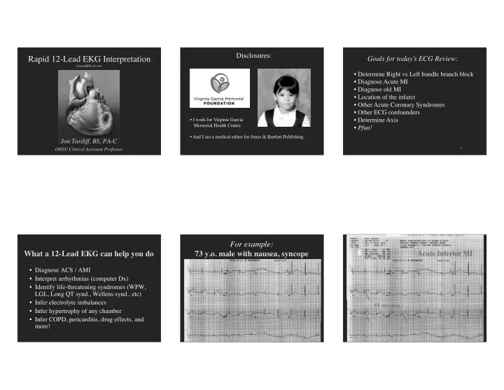
For example: 73 y.o. male with nausea, syncope
6 6
Acute Inferior MI
ST elevation