A Rare Nasopharyngeal Presentation The Author(s) 2020 Article reuse - - PDF document

a rare nasopharyngeal presentation
SMART_READER_LITE
LIVE PREVIEW

A Rare Nasopharyngeal Presentation The Author(s) 2020 Article reuse - - PDF document

Case Report Ear, Nose & Throat Journal 14 A Rare Nasopharyngeal Presentation The Author(s) 2020 Article reuse guidelines: sagepub.com/journals-permissions of Amyloidosis DOI: 10.1177/0145561320914432 journals.sagepub.com/home/ear


slide-1
SLIDE 1

Case Report

A Rare Nasopharyngeal Presentation

  • f Amyloidosis

Erika Crosetti, MD, PhD1, Andrea Manca, MD1 , Elena Maldi, MD2, and Giovanni Succo, MD1,3

Abstract Amyloidosis is a heterogeneous group of diseases characterized by the extracellular deposition of insoluble proteins whose pathogenesis is not yet fully understood. The deposition of amyloid proteins can be systemic or localized, idiopathic or related to systemic diseases, mostly multiple myeloma or chronic inflammatory diseases. Localized head and neck amyloidosis is a rare entity, mainly involving the larynx. Given the rarity of the disease and the absence of a lasting follow-up protocol, there is no standard treatment defined for localized amyloidosis. We report a rare case of localized nasopharyngeal amyloidosis, treated with complete transoral resection and confirmed by histological examination. Keywords amyloidosis, nasopharynx, head and neck diseases, localized

Introduction

Amyloidosis is a heterogeneous group of diseases character- ized by the deposition of insoluble proteins in the extracellular

  • space. The deposition of amyloid proteins can be systemic or

localized.1,2 The systemic form can be idiopathic or related to systemic disease, mainly multiple myeloma or chronic inflammatory

  • diseases. Localized nasopharyngeal amyloidosis is an

extremely rare entity with few cases reported in the literature. Amyloidosis can be suspected based on clinical signs and imaging studies but the definitive diagnosis requires tissue biopsy, analyzed using Congo red staining combined with polar- ized light and possibly immunohistochemical characterization of the proteins. There is no specific cure for amyloidosis and treat- ment depends on the form of the disease, the organs affected, and the underlying condition. We describe a case of an 80-year-old woman with a primary localized nasopharyngeal amyloidosis.

Case Report

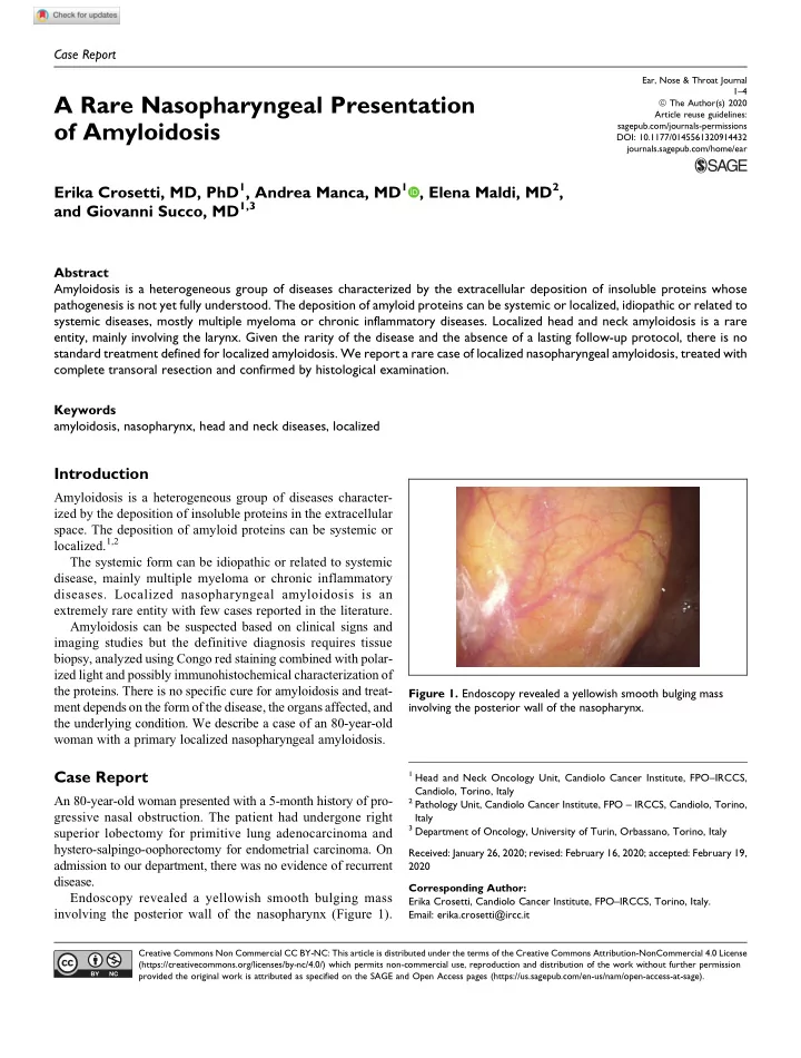
An 80-year-old woman presented with a 5-month history of pro- gressive nasal obstruction. The patient had undergone right superior lobectomy for primitive lung adenocarcinoma and hystero-salpingo-oophorectomy for endometrial carcinoma. On admission to our department, there was no evidence of recurrent disease. Endoscopy revealed a yellowish smooth bulging mass involving the posterior wall of the nasopharynx (Figure 1).

1 Head and Neck Oncology Unit, Candiolo Cancer Institute, FPO–IRCCS,

Candiolo, Torino, Italy

2 Pathology Unit, Candiolo Cancer Institute, FPO – IRCCS, Candiolo, Torino,

Italy

3 Department of Oncology, University of Turin, Orbassano, Torino, Italy

Received: January 26, 2020; revised: February 16, 2020; accepted: February 19, 2020 Corresponding Author: Erika Crosetti, Candiolo Cancer Institute, FPO–IRCCS, Torino, Italy. Email: erika.crosetti@ircc.it

Figure 1. Endoscopy revealed a yellowish smooth bulging mass involving the posterior wall of the nasopharynx.

Ear, Nose & Throat Journal 1–4

ª The Author(s) 2020

Article reuse guidelines: sagepub.com/journals-permissions DOI: 10.1177/0145561320914432 journals.sagepub.com/home/ear Creative Commons Non Commercial CC BY-NC: This article is distributed under the terms of the Creative Commons Attribution-NonCommercial 4.0 License (https://creativecommons.org/licenses/by-nc/4.0/) which permits non-commercial use, reproduction and distribution of the work without further permission provided the original work is attributed as specified on the SAGE and Open Access pages (https://us.sagepub.com/en-us/nam/open-access-at-sage).

slide-2
SLIDE 2

Maxillofacial magnetic resonance imaging showed a submuco- sal mass (26 14 43 mm) involving the posterior wall of the nasopharynx, characterized by polycyclic margins, homoge- neous contrast enhancement, isointensity with muscles in T1 and T2 acquisitions, and hyperintensity in Short-TI Inversion Recovery images, without infiltrative aspects (Figure 2). A total body computed tomography (CT) scan showed no other solid lesions. The nasopharyngeal mass had inhomogeneous contrast enhancement and was accompanied by 2 retropharyn- geal nodes. Considering the radiologic features, the poor vasculariza- tion and the distance from the large neck vessels, a complete transoral resection was carried out. The surgical specimen was routinely processed. The postoperative period was uneventful. Microscopic examination revealed a submucosal deposi- tion of cloudy, ill-defined pinkish material highly suspicious for amyloid deposition (Figure 3). Specific Congo red stain- ing was performed and revealed apple-green birefringence under polarized light (Figure 4). Multiple attempts were made to further characterize the type of amyloid proteins using immunohistochemistry but with negative results, thus a diag- nosis of nasopharyngeal amyloid deposition, unspecific type, was made. Consequently, a complete work-up was performed, ruling

  • ut systemic involvement and conditions possibly related to a

secondary form of amyloidosis. A diagnosis of localized naso- pharyngeal amyloidosis was then postulated. Twenty-four months after surgery, our patient is in good condition and disease-free, undergoing regular clinical endoscopic and radi-

  • logical follow-up.

Discussion

Amyloidosis is a rare clinical entity, characterized by the extra- cellular deposition of proteinaceous material comprised of var- ious abnormal proteins known as amyloid fibrils.3 Amyloid deposits result from a soluble precursor protein which is mis- folded resulting in the deposition of insoluble amyloid fibrils.4 Amyloidosis is defined as localized if the amyloid deposition is limited to the tissue where its precursor protein is produced, and as systemic if the precursor protein is produced in a certain part of the body and thereafter transported to the site where the amyloid is found.5 The modern classification of amyloidosis uses an abbrevia- tion of the predominant protein, preceded by the letter A. Among the systemic forms, there are immunoglobulin light chain amyloidosis (AL), acute phase protein amyloidosis (AA), dialysis-related amyloidosis (Ab2M, b2 microglobulin), and age-related amyloidosis (ATTR, transthyretin). Microscopically, amyloidosis is characterized by deposition

  • f ill-defined pink material. In the localized form, a plasma cell

infiltrate is described.6 Congo red staining confirms the amy- loid nature with a classic apple-green birefringence. Immuno- histochemistry with commercially available antibodies should be performed in order to characterize the protein component. Clinical presentation depends on the organs affected by the

  • deposit. The kidneys are frequently involved in AL amyloido-

sis and this can lead to kidney failure; the same loss of function can affect the heart or lungs. In the head and neck region, the

Figure 2. Maxillofacial magnetic resonance imaging showed a submucosal mass (26 14 43 mm) involving the posterior wall of the nasopharynx, characterized by polycyclic margins, homogeneous contrast enhancement, isointensity with muscles in T1 and T2 acquisitions, and hyperintensity in Short-TI Inversion Recovery (STIR) images, without infiltrative aspects. Figure 3. Microscopic examination revealed a submucosal deposition

  • f cloudy, ill-defined pinkish material highly suspicious for amyloid

deposition.

2 Ear, Nose & Throat Journal

slide-3
SLIDE 3

most frequent symptoms are hoarseness and dyspnea in laryn- geal forms, or macroglossia in cases with oral cavity involvement.6 The prognosis of the AA form depends on the condition responsible for the amyloidosis itself. The AL amyloidosis can be treated with chemotherapy and stem cell transplantation; on the other hand, the AA form is treated aiming to cure the underlying disease. Localized amyloidosis in the head and neck region is rare. Most cases reported have been described in the larynx (60%), oropharynx (23%), trachea (9%), orbit (4%), and nasopharynx (3%).7-9 Nasopharyngeal amyloidosis is extremely rare. In their review in 2007, Panda et al described only 13 cases in over 70 years of activity (1935-2007).10 Lately, there has been an increase in the number of reported cases, perhaps due to improvements in endoscopic and radiologic diagnostic tools.11,12 Patients with nasopharyngeal amyloidosis mainly complain

  • f nasal obstruction, epistaxis, posterior rhinorrhea, and hear-

ing impairment.13 Endoscopy generally shows an irregular, heterogeneously yellowish, smooth bulging mass.14 On CT scan, these lesions usually appear as a well-defined, homoge- nous, smooth soft tissue mass without evidence of bony ero- sion.15 It has also been reported to mimic slow-growing tumors and sometimes to appear as densely calcified lesions.16 Imaging can help to define the extent of the disease but there are no characteristic imaging findings, so the diagnosis cannot be made without a biopsy or excision.17,18 Diagnosis is then confirmed by histopathology, with characteristic apple-green birefringence on microscopy using Congo red staining com- bined with polarized light. Given the rarity of the disease and the absence of a lasting follow-up protocol, no standard treatment has been defined for localized amyloidosis. Treatment of local disease is by com- plete excision when this is possible without significant anato- mical or functional sequelae, followed by routine surveillance for recurrence. Clinical follow-up is by endoscopic examina- tion, supported by imaging when a recurrence is suspected, based on signs and symptoms. At present, there is no evidence in the literature suggesting a progression from nontreated localized head and neck amyloidosis to systemic disease.19 Thus, at present, there is an ongoing debate between surgical excision, preferred when the disease causes morbidity, and a wait-and-see approach. Declaration of Conflicting Interests

The author(s) declared no potential conflicts of interest with respect to the research, authorship, and/or publication of this article.

Funding

The author(s) disclosed receipt of the following financial support for the research, authorship, and/or publication of this article: This manu- script was funded by the Regione Piemonte AD FUNCTIONEM (years 2015–2018); FPRC 5xmille 2016 Ministero della Salute Pro- getto ARDITE; Fondi Ricerca Corrente 2018, Ministero della Salute.

ORCID iD

Andrea Manca https://orcid.org/0000-0003-4267-8066

References

  • 1. Westermark P, Benson MD, Buxbaum JN, et al. Amyloid: toward

terminology clarification: Report from the Nomenclature Com- mittee of the International Society of Amyloidosis. Amyloid. 2005;12(1):1-4.

  • 2. Werner BC, Shen FH, Shimer AL. Primary cervical amyloidoma:

a case report and review of the literature. Spine J. 2013;13(10): e1-7.

  • 3. Sipe JD, Benson MD, Buxbaum JN, et al. Amyloid fibril proteins

and amyloidosis: chemical identification and clinical classifica-

  • tion. International Society of Amyloidosis 2016 Nomenclature
  • Guidelines. Amyloid. 2016;23(4):209-213.
  • 4. Nienhuis HLA, Bijzet J, Hazenberg BPC. The prevalence and

management of systemic amyloidosis in Western countries. Kid- ney Dis. 2016;2(1):10-19.

  • 5. Sipe JD, Benson MD, Buxbaum JN, et al. Nomenclature 2014:

amyloid fibril proteins and clinical classification of the amyloi-

  • dosis. Amyloid. 2014;21:221-224.
  • 6. Penner CR, Muller S. Head and neck amyloidosis: a clinicopatho-

logic study of 15 cases. Oral Oncol. 2006;42(4):421-429. Figure 4. Congo red staining (left, clear light, OM 40) revealed apple-green deposits under polarized light (right, OM 40).

Crosetti et al 3

slide-4
SLIDE 4
  • 7. Glenner G. Amyloid deposits and amyloidosis: the beta-

fibrilloses (second of two parts). N Engl J Med. 1980;302(24): 1333-1343.

  • 8. Glenner G. Amyloid deposits and amyloidosis: the beta-

fibrilloses (first of two parts). N Engl J Med. 1980;302(23): 1283-1292.

  • 9. Scott PP, Scott WW, Siegelman SS. Amyloidosis: an overview.

Semin Roentgenol. 1986;21(2):103-112.

  • 10. Panda NK, Saravanan K, Purushotaman GP, et al. Localized amy-

loidosis masquerading as nasopharyngeal tumor: a review. Am J

  • Otolaryngol. 2007;28(3):208-211.
  • 11. Sakagiannis G, Giotakis E, Thompson LDR. Localized

nasopharyngeal amyloidosis: a clinicopathologic series of 7 cases with a literature review. Head Neck Pathol. 2018;12(4):542-547. http://dx.doi.org/10.1007/s12105-017- 0880-4.

  • 12. Yoshida A, Borkar S, Singh B, et al. Incidental detection of con-

current extramedullary plasmacytoma and amyloidoma of the nasopharynx on [18F]fluorodeoxyglucose positron emission tomography/computed tomography. J Clin Oncol. 2008;26(35): 5817-5819.

  • 13. Durbec M, Ambrun A, Barnoud R, et al. Localized nasopharyngeal
  • amyloidosis. Eur Ann Otorhinolaryngol Head Neck Dis. 2012;

129(3):160-162. http://dx.doi.org/10.1016/S1726-4901(10)70119-2.

  • 14. Chen Y-S, Li W-Y, Ho C-Y. Localized nasopharyngeal amyloi-
  • dosis. J Chin Med Assoc. 2010;73(10):549-552.
  • 15. Harirchian S, Mirani N, Baredes S. Amyloidosis of the nasophar-

ynx and nasal cavity presenting as severe epistaxis and sphenoid sinus dehiscence. Laryngoscope. 2011;121(4):S73-S73. 10.1002/ lary.21943.

  • 16. Parmar H, Rath T, Castillo M, et al. Imaging of focal amyloid

depositions in the head, neck, and spine: amyloidoma. Am J Neu-

  • roradiol. 2010;31(7):1165-1170.
  • 17. Gean-Marton AD, Kirsch CFE, Vezina LG, et al. Focal amyloi-

dosis of the head and neck: evaluation with CT and MR imaging.

  • Radiology. 1991;181(2):521-525.
  • 18. Urban BA, Fishman EK, Goldman SM, et al. CT evaluation of

amyloidosis: spectrum of disease. Radiographics. 1993;13(6): 1295-1308.

  • 19. Nandapalan V, Jones TM, Morar P, et al. Localized amyloidosis
  • f the parotid gland: a case report and review of the localized

amyloidosis of the head and neck. Head Neck. 1998;20(1):73-78.

4 Ear, Nose & Throat Journal