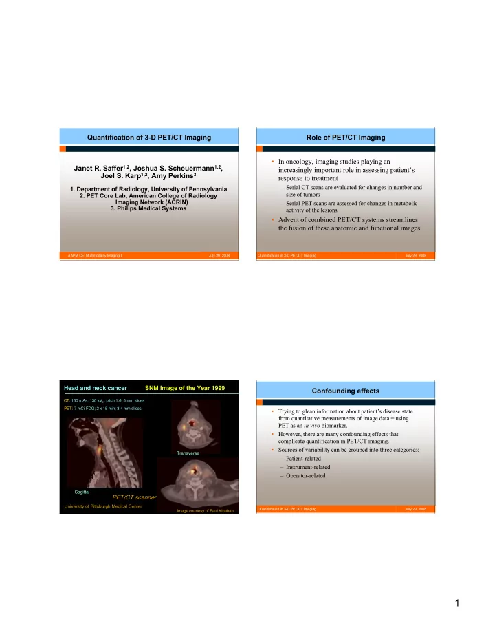
SLIDE 7 7
July 29, 2008 Quantification in 3-D PET/CT Imaging
The Netherlands Protocol
- Protocol for standardization of quantitative FDG whole body PET
studies
- Based on standardization of:
1. patient preparation 2. matching of scan statistics by prescribing dosage as function of patient weight, scan time per bed position, percentage of bed overlap and image acquisition mode (2D or 3D) 3. matching of image resolution by prescribing reconstruction settings for each type of scanner 4. matching of data analysis procedure by defining volume of interest methods and SUV calculations 5. a multi-center QC procedure using a 20 cm diameter phantom for verification of scanner calibration and the NEMA NU 2 2001 Image Quality phantom for verification of activity concentration recoveries (i.e. verification of image resolution and reconstruction convergence).
Boellaard R, Oyen W, Hoekstra C, et al. The Netherlands protocol for standardization of FDG whole body PET studies in multi-center trials (NEDPAS), J Nucl Med (2008); 49 (Supplement 1):106P. July 29, 2008 Quantification in 3-D PET/CT Imaging
AAPM Task Groups on Related Topics
- AAPM Task Group 126 – “PET/CT Acceptance Testing and Quality
Assurance”, Chair: Osama Mawlawi (MD Anderson Cancer Ctr).
- AAPM Task Group 145 - “Quantitative Imaging Initiative: Quantitative
PET/CT Imaging Joint AAPM/SNM Committee”, Chair: Paul Kinahan (U Wash). Goal: to develop procedures and tools that allow proper pooling of results from multicenter trials to achieve/increase statistical and clinical significance.
- AAPM Task Group 174 - “Utilization of 18F-Fluorodeoxyglucose
Positron Emission Tomography (FDG-PET) in Radiation Therapy”, Chair: Shiva Das (Duke). Goal: to recommend guidelines/protocols for consistent imaging, consistent treatment planning and consistent treatment assessment using FDG-PET in radiotherapy.
July 29, 2008 Quantification in 3-D PET/CT Imaging
Acknowledgements ACRIN PET Core Lab Key Personnel:
- PET/Nuclear Medicine Steering Committee
Chair/Medical Director: Barry Siegel, MD, Washington University
- Technical Supervisor: Anthony Levering, RT
July 29, 2008 Quantification in 3-D PET/CT Imaging
References (in chronological order)
1) Lowe VJ, Delong DM, Hoffman JM, Coleman RE. Optimum scanning protocol for FDG-PET evaluation of pulmonary malignancy. J Nucl Med (1995); 36:883-87. 2) Young H, Baum R, Cremerius U, et al. Measurement of clinical and subclinical tumour response using [18F]-fluorodeoxyglucose and Positron Emission Tomography: review and 1999 EORTC recommendations. Eur J Cancer (1999); 35(13):1773-1782. 3) Therasse P, Arbuck SG, Eisenhauer EA, et al. New Guidelines to Evaluate Response to Treatment in Solid Tumors. J NCI (2000); 92(3):205-216. 4) Nakamoto Y, Osman M, Cohade C, Marshall LT, Links JM, Kohlmyer S, Wahl RL. PET/CT: comparison of quantitative tracer uptake between germanium and CT transmission attenuation-corrected images. J Nucl Med (2002); 43(9):1137-43. 5) Weber WA. Use of PET for monitoring cancer therapy and for predicting outcome. J Nucl Med (2005); 46(6):983-995. 6) Coleman RE, Delbeke D, Guiberteau MJ, et al. Concurrent PET/CT with an integrated imaging system: Intersociety dialogue from the joint working group of the American College of Radiology, the Society of Nuclear Medicine, and the Society of Computed Body Tomography and Magnetic Resonance. J Nucl Med (2005); 46(7):1225-1239. 7) Delbeke D, Coleman RE, Guiberteau MJ, et al. Procedure guideline for tumor imaging with 18F-FDG PET/CT 1.0, J Nucl Med (2006); 47(5):885-95. Page 1 of 2