
SLIDE 1
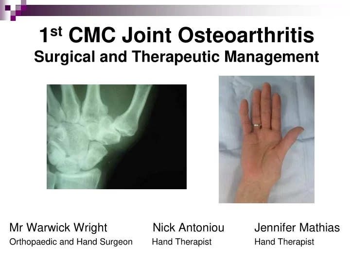
1st CMC Joint Osteoarthritis
Surgical and Therapeutic Management
Mr Warwick Wright Nick Antoniou Jennifer Mathias
Orthopaedic and Hand Surgeon Hand Therapist Hand Therapist
1 st CMC Joint Osteoarthritis Surgical and Therapeutic Management - - PowerPoint PPT Presentation
1 st CMC Joint Osteoarthritis Surgical and Therapeutic Management Mr Warwick Wright Nick Antoniou Jennifer Mathias Orthopaedic and Hand Surgeon Hand Therapist Hand Therapist 1 st CMC joint OA in brief Most common hand OA (after DIPJs)
Orthopaedic and Hand Surgeon Hand Therapist Hand Therapist
Most common hand OA (after
6:1 Female:Male (high as 10-15:1) Major cause of thumb & hand
Ligamentous Laxity
AOL becomes lax with adjacent palmar degeneration of trapezium (or
dorsoradial laxity and adjacent dorsal degeneration) Joint Impingement
Degeneration secondary to joint impingement during functional pinch
(lateral pinch)
High contact stresses through pinch initiate and/or exacerbate OA
Moulton et al 2001, Bettinger et al 2000, Imaeda et al 1999 Kovler et al 2004, Koff et al 2003, Ateshian et al 1995
Thumb MC rests in plane perpendicular to palm – enabling functional pinch 3 Planes of movement
Flexion / Extension (RA) Abduction / Adduction Opposition / Retropulsion
Bi concave / convex (imperfect)
Shallow (bony congruity / stability poor)
Stability largely from ligaments (16) and muscle tendon units (9)
Large contact forces at CMCJ from tip pinch (factor of x 6 - 24 at CMCJ)
Degeneration at volar-ulnar quadrant
Anterior Oblique Ligament (AOL) the major (static) stabilising structure – limits dorso- radial translation of the MC on the trapezium in pinch
Dorso-Radial Ligament (DRL) is now considered to be just as important a stabiliser – taut during MC dorsoradial subluxing forces
AOL attenuation causes degeneration to the adjacent volar / ulnar aspect of the trapezium
Lateral (key) pinch causes concentrated forces in same zone
Bettinger 2001,
Maximal contact area between
Trapezium and Metacarpal (53%) during opposition (abduction, flexion and pronation)
Ligaments taut in this position Most stable “close packed” position is
“screw-home-torque position”
Neumann and Bielefield 2003
Strong thumb adductor (flexor and
Transverse and Oblique heads Strong in lateral (key) pinch Significant contributor to thumb OA
Serves as an important
CMCJ stabiliser (counteracts action of AP)
Aberrent accesory tendons
Trapezium)
? minimises OA prevalence
as pull of APL on both Metacarpal and Trapezium causes concurrent pulling (less shear)
No correlation found
Roush et al 2005, Schultz et al 2002, Roh et al 2002
MCPJ hyperextension
accentuates deformity Adduction Contracture
Dorsoradial Subluxation of MC
IPJ flexion
CMCJ instability causative of MCPJ
deformity but divergent theory of MCPJ being causative
MCPJ flexion unloads volar surface of
trapezium (30° causes 60% dorsal shift
CMCJ congruence facilitated
in MCPJ position of 30° flexion
Ambruster & Tan 2008, Moulton et al 2001, Johnson et al 1996
X-rays Patient history of pain and
Clinical assessment
shoulder sign / deformity palpation grind test
Pain Function
Thumb AROM
Strength (Pinch and Grip)
Pain (5 items)
(at rest, gripping, lifting, turning, squeezing)
Stiffness (1 item)
(on waking)
Physical Function (9 items)
(turning taps/faucets on, turning a round doorknob or handle, doing up buttons, fastening jewellery, opening a new jar, carrying a full pot with
wringing out wash cloths)
Rest Splinting Heat Exercise NSAIDS CSIs Activity Modification and JPE Assistive devices
Maximise (painfree) functional ROM Maximise functional strength and
endurance
Maintain stability of the CMCJ Reduce pain Avoid fixed deformities
Kjeken 2011, Neumann and Bielefeld 2003, Felson 2000
Aims
Encourage joint motion and tissue elasticity
(cartilage nutrition and joint lubrication)
Restore web space Maintain functional strength for pinch and grasp Condition muscles to absorb damaging impact loads
Principles
A/PROM (all planes) as well as conventional strengthening for
functional pinch and grasp
Felson 2000, Neumann and Bielefeld 2003
A/PROM
CMCJ Abduction/Adduction/
Flexion/Extension/ Opposition/Retropulsion/ Composite
“Place and Hold”
Resistance
Pinch (Lateral / Tip) Grip Isometrics / Putty / etc
Garfinkel et al 1994, Lefler & Armstrong 2004, Wajon & Ada 2005, Rogers & Wilder 2009, Boustedt et al 2009
Kjeken et al (2011) may reduce pain and increase ROM and strength Ye et al (2011) exercise has no effect on hand pain / dysfunction although may be able to improve hand strength Valdes and Marik (2010) moderate evidence to support hand exercises for increasing grip, improving function, ROM and pain reduction Not specific to thumb OA / thumb exercises
Sackett et al (2000) Scale
Rogers & Wilder (2009)
Study Type: Crossover trial (level 2b), n=46 with hand OA in 1 joint Program: 16 week program for each (16 week washout in between) Exercise vs Sham (hand cream daily) Exercises: x 1 daily, 10 reps 20 reps over 16 weeks AROM - table top / hook / full fist / opposition all digits / finger spread / thumb flexion Strengthening - Theraband Ball - grip / lat pinch / tip pinch High attrition rate – 40% (n=30), mostly in exercise group
No change in AUSCAN or dexterity but significant improvement in grip and key pinch
Lefler & Armstrong (2004) Study Type: RCT (level 1b), n=19 with hand OA in 1 joint Program: 6 week program of strengthening x 3 p/week –
Exercises: (1) Rice grabs, (2) 5 finger pinch grip lifting (sand bags) / wrist rolls with PVC pipe attached to 250g sand bag Sig improvement in grip and ROM but not pain or pinch strength
Wajon & Ada (2005) Study Type: RCT (level 2b), n=40 with thumb OA Program: 4 week program, 5-10 reps (and increasing as pain allows) x 3 p/day Exercises: Thumb abduction against gravity (and thumb strap splint) vs foam block finger tip pinch (and short opponens splint) High bias risk – differing splints (major confounding variable) No significant difference between the 2 programs
promote muscular (dynamic) stability of the CMCJ maintain first web space (limit adduction deformity)
APB – small & weak but positions thumb for pinch and palmarly abducts
and pronates (screwing action) – puts CMC joint in maximal stability (bony and ligamentous)
APL – strong muscle that abducts thumb and pulls MC radially. Opposes
the powerful adductors of the thumb and limits dorso-radial collapse of MC and narrowing of 1st web space.
EPL - not desirable as acts as adductor. Use sparingly to maintain
flexibility in absence of established deformity
Wajon & Ada 2005, Neumann and Bielefeld 2003, Poole & Pellegrini 2001
Neumann and Bielefeld 2003, Smutz et al 1998
Isometrics Rubber band Theraputty
early stages, as later can destabilising and contribute to
subluxation (eg. EPL)
painfree (non-inflammed) state close packed position or end range active or resisted (isometrics less traumatic alternative) pain following performance < 2 hours acceptable
Poole & Pellegrini 2001
Based on Jan Albrecht’s approach “Caring for the painful thumb;
more than a splint…”
Use of thumb muscles during function to stabilise the CMCJ to
reduce / prevent subluxating shear forces.
Functional kinematic approach superior to traditional strengthening Entire set of muscles around joint to centralise / restore function Concept of “pertubation” training
In Summary:
Indicated for painful thumbs (irrespective of stage / pathology) Restoration of thumb web space Re-education of intrinsics / extrinsics (esp FDI, OP and abductors
and extensors)
Joint mobilisation techniques Strengthening to reinforce muscle patterns for joint stability
(restore order & strength of muscle recruitment through full ROM)
Combined interventions (Splintage / JPE / Adaptive equip) Order of intervention a clinical decision
“lateral thenar muscle” distal / ulnar forces of FDI
counteract the dorso-radial forces of lateral pinch and grip
causes distraction rather
than compression of CMCJ
Brand & Hollister 1993
Study Retrospective, n=35, (Level 4) Unstructured JPE / splintage intervention QuickDASH scores x 2 (initial / last) Results Pain score reduced 17.9% (significant) Function score improved 19.3% (significant) (DASH MCID of 15%) Positive results achieved at 2nd visit over 6 weeks
Poor study design, retrospective and confounders (splintage / JPE) Radiographic subluxation change not measured (only DASH)
Splinting: Pain -
At rest (with / out splint) During activity (with / out splint)
Splint weaned when fx pinch painfree Exercises:
Opposition AP myofascial release
(contract / relax)
“Web space comparison”
CMC joint extension
Distraction of joint using other
Dorsal subluxation reduction
Retroposition – hold 1-3 mins
FDI (AROM resistance)
APB / EPB / OP “C” position Oppositional pinch P+H
Proprioceptive taping
As needed
Based on the American College of Sports Medicine (ACSM)
recommendations for “developing muscular strength and flexibility in older frail adults”
Explored exercise dosage (not specifically goal of exercise) Dosage parameters
(load, reps/set, sets, sets/day, duration, max or painfree)
General Principles
Strengthening should be 40-50% of 1 rep max effort. LP strengthening avoided in advanced OA (III and IV) (contributes
to joint subluxation and pain)
Given x 6-24 factor of load at CMC, consider these loads when
performing pinch and grip exercise
Painfree Principle Pain to not exceed > 2 hrs after activity Heat or low intensity aerobic exercise beforehand Minimum 12 weeks
Composite thumb flexion to base
Abduction + Opposition Isolated IP and MCP joint flexion CMC extension (watch MCPJ
hyperextn)
Principles
2-4 reps and > 2-3 days p/week – but daily is best Stretch to point of tightness or slight discomfort
(+/- assisted stretch of 10-30 secs)
10-30 secs hold static stretch but 30-60 secs in older persons Heat beforehand
thumb extension and abduction against
resistance (rubber band, velcroboard, putty)
Isometrics Pinch (if appropriate) using putty / pegs Grip using putty / hand grippers / foam
wedge squeeze
All thenar intrinsics (except AP), extrinisc thumb extensors, abductors and wrist extensors
Principles
Lateral (key) pinch avoided in advanced OA or presence of
instability / deformity
Each muscle group trained x 2-3 p/week 10-15 reps x 1 set (minimum) with 2-3 mins rest between > 48 hrs rest between sessions
Minimal evidence available overall and especially of thumb Dynamic stability of thumb CMC joint through targeted muscle
strengthening considered to be important – no evidence as yet to prove this
Avoidance of AP strengthening Avoidance of LP strengthening
Some guidelines for dosage now established
Flexibility, daily performance and to point of stretch discomfort Strengthening, x 2-3 per week – painfree principle At least 12 weeks (?indefinately)
Minimise deformities Decrease pain Provide support for increased function Decrease stress to the joints Decrease inflammation Assist with joint stability Increase stability Reduce mechanical stress that may cause instability
Prevent first webspace contracture
Gives the therapist time to develop a therapeutic rapport with the pt Assess the severity of the symptoms Prevent adduction of the metacarpal head into the palm & dorsoradial subluxation of the MC base on the trapezium
Splint design features Type of Splint Custom / Prefabricated Material Rigid / Soft / Combination Joints Immobilised CMCJ / wrist / MCPJ Wearing regime Wearing regime Continuously (rest & function) Vs Intermittently (function) Goal of splint
webspace
unload the palmer compartment of the CMC joint Type of splint / material used / wearing regime
Design
subjects Type of splint Splint wear Length of study Results
Low-quality RCT (2b) 40 patients Thermoplastic splint to stabilise the CMC, IP joint free, functional position Rx group – Splint for ADL’s for 180 days Control group – Splint for the Ax’s, then ADL’s for 90 days. 180 days No improvement in function in both groups. No change in grip strength in both groups. Pinch strength reduced in both groups following splinting. No change in dexterity with both groups. Pain reduced in the treatment group (from the first evaluation at 45 days) and the control group once they commenced wearing the splint at day 90.
Outcome measures
VAS pain scale DASH questionnaire Grip strength (Jamar) Pinch strength (pinch guage) UL dexterity test
Gomes Carreira, A, Jones A and Natour J. Assessment of the Effectiveness of a Functional Splint for Osteoarthritis of the Trapeziometacarpal Joint of the Dominant Hand: A Randomized Controlled Study. J Rehabil Med. 2010; 42: 469-474
Weiss S, LaStayo P, Mills A, Bramlet D. Prospective Analysis Of Splinting The First Carpometacarpal Joint: An Objective, Subjective And Radiographic
26 Design
subjects Type of splint Splint wear Length of study Results
Cross over 2b 26 subjects 1) CMC splint 2) CMC splint and MCP splint Wear splints whenever symptoms are felt (day or night) 2 weeks Each splint was used for
Both splint groups had a reduction of pain, but there was no significant difference between the 2 groups. No change in pinch strength or in reducing pain during pinch with both groups. Both splints reduce CMC subluxation. Pts with grades 1 and 2 had better stabilisation of the first CMC joint with each splint than did pts with grade 3 or 4. CMC splint was the preferred splint
Outcome measures
VAS pain scale Tip pinch guage CMC subluxation (X-rays) ADL self rated scale
Weiss, S, LaStayo P, Mills A and Bramlet D. Splinting the Degenerative Basal Joint: Custom-made or Prefabricated Neoprene? Journal of Hand Therapy; Oct-Dec 2004: 17,4: 401-406 Design
subjects Type of splint Splint wear Length of study Results
Cross over (2b) 25 subjects 1) Custom-made short opponens thermoplastic splint 2) Prefabricated neoprene splint Pts instructed to wear splint whenever they felt symptoms (day or night) Wear splint 1 for
swap to splint 2 for one week. 2 weeks Each splint was used for
Thumb pain decreased after wearing each of the splints. Pain was significantly less when wearing neoprene splint. Pain at rest and pain during pinch improved more significantly in the neoprene group compared to thermoplastic group. Tip pinch strength (splint on) improved more significantly in the neoprene group. Neoprene group more satisfied with the splint vs thermoplastic group The CMC joint subluxation was more significantly reduced in the thermoplastic group compared to the neoprene group.
Outcome measures
VAS pain scale CMC subluxation (X-rays) Pinch strength (with pinch meter) VAS splint satisfaction Self rated scale of ADL’s
Rannou F, Dimet J, Boutron I, Baron G, Fayed F, et al. Splint for Base-of-Thumb Osteoarthritis. Annals of Internal Medicine. 2009, 150: 10: 661-669 Design
subjects Type of splint Splint wear Length of study Results
RCT (1b) 112 subjects 1) Custom-made splint 2) Usual care Wear at night only One year At 1 month no difference between the 2 groups in all areas measured. At 12 months there was a significant improvement in pain and function in the splinted group compared to the control group. The splint had no effect on the radiographic progression of OA.
Outcome measures
VAS pain scale VAS pts perceived disability Cochin Hand Functional Scale Pt global assessment Pinch strength (dynamometer) ROM (kapandji score) X-rays
Design
subjects Type of splint Splint wear Length of study Results
2b (RCT) 40 1) Treatment group: Thumb strap splint and abduction ex 2) Control group: Short opponens splint and pinch grip ex Splint full time 2 weeks of splinting alone (either thumb strap
Then exercises were introduced at 2 weeks, (and splinting continued) At week 2 and week 6, no differences in VAS scores, tip pinch strength or Sollerman Test of Hand Function scores between the 2 groups. However, both groups improved in regards to pain, tip pinch strength and function.
Outcome measures
VAS pain scale Pinch strength (pinch guage) Sollermann hand function test
Wajon, A and Ada L. No Difference Between Two Splint And Exercise Regimes For People With Osteoarthritis Of The Thumb: A Randomised Controlled Trial. Australian Journal of Physiotherapy. 2005 51: 245-249
Swigart CR, Eaton RG, Glickel SZ, Johnson
The First Carpometacarpal Joint. Journal of Hand Surgery Am. 1999; 24; 86-91 Design
subjects Type of splint Splint wear Length of study Results
3 (Cohort study) 130 subjects Long opponens splint incl. wrist Full time wear for 3-4 weeks, then weaning period of 3-4 weeks. 6 months Reduction in the severity of symptoms, allowing function without significant pain.
Outcome measures
X-ray to Ax stage Questionnaire
Buurke JH, Grady JH, de Vries J, Baten CT. Usability Of Thenar Eminence Orthoses: Report Of A Comparative Study. Clin Rehabil. 1999: 13: 288-94 Design
subjects Type of splint Splint wear Length of study Results
Randomised cross over (2b) 10 subjects 1) Semi-rigid 2) Firm elastic 3) Supple elastic No instruction 12 weeks Each splint used for 4 weeks Better hand function in gripping with soft splint and better tolerated No difference between the 3 groups with pain.
Outcome measures
VAS pain scale Pinch test (guage) Hand function in hand grips (Green test) VAS hand function VAS cosmesis of splint
Sillem H, Backman C, Miller W, Li L. Comparison Of Two Carpometacarpal Stabilizing Splints For Individuals With Thumb
Design
subjects Type of splint Splint wear Length of study Results
2b (cross over trial) 56 subjects 1) Hybrid splint 2) Comfort cool CMC splint Wear in the day when symptomatic and at night as desired 9 weeks 4 weeks wearing one splint One week off 4 weeks wearing the
Comfort cool was the preferred splint Hybrid splint group had a significant reduction in pain than those in comfort cool group Both groups reported improved hand function
Outcome measures
AUSCAN Grip strength (Jamar) Pinch strength (pinch meter) Scale re: preference with fit, appearance, convenience, and durability
2000)
2000)
(Poole & Pelligrini 2000)
With STT joint With MCP joint With IP joint CMC joint
Thermoplastic Neoprene with thermoplastic Custom made Neoprene Off the shelf Day Night Day & Night
Heavy tasks Full-time Off for sedentary tasks
Maintain web space stretch Involve MCP to unload CMC
Wearing Regime Joint affected
Stabilise the base of the 1st MC during pinch
Material
Respect Pain Balance rest and activity Exercise in a pain free range Reduce the effort and force Use larger /stronger joints Avoid positions of deformity
Beasley J. Osteoarthritis and Rheumatoid Arthritis: Conservative Therapeutic Management. Journal of Hand Therapy. April – June 2012
Susan Michlovitz, PhD, PT, et al. Continuous Low-Level Heat Wrap Therapy Is Effective For Treating Wrist Pain. Archives of Physical Medicine and Rehabilitation. September 2004. Vol. 85. No. 9. Pp. 1409-1416.
The authors conclude that low-level continuous heat wraps can help in the treatment
Blood helps remove cells of inflammation in the area of tissue injury. The collagen tissue and muscles then become more flexible.
In the Cochrane review 2010 = there is weak evidence to support the use of paraffin wax for pain reduction, ROM and improved hand function Moderate evidence to support the use of continuous heat packs for pain reduction and increased grip strength
Ligament Reconstruction (LR) MC osteotomy TMCJ arthrodesis Denervation TMCJ replacement Trapeziectomy +/- LR or TI or LRTI Trapeziectomy (complete/partial) + interpositional arthroplasty
Interposition = any material / tissue interposed between the thumb MC and scaphoid
Arthroplasty = any procedure where the joint is reconstructed (partially or completely) Trapeziectomy – any procedure where the complete or partial removal of the trapezium bone is performed Partial trapeziectomy = Hemiarthroplasty / Resurfacing arthroplasty Ligament reconstruction (LR) = reconstructing the AOL, with tendon graft – not always performed Suspensionplasty = technical variation using the APL tendon to suspend the first MC through its base and to the IF MC to minimise collapse during pinch
So which procedure is best? Cochrane Review: Wajon et al, (2009), Surgery for Osteoarthritis of the Thumb
“…although no one procedure produces greater benefit in terms of pain and physical function, there was insufficient evidence to be conclusive. Trapeziectomy has fewer complications than trapeziectomy with LRTI.”
Varies, depending on procedure, surgeon and therapist Main considerations/parameters:
Period of immobilisation (1-6 weeks) Spica cast removal timing (1-6 weeks) Short or Long opponens splint Position of thumb in splint (encourage/discourage fx pinch) Splint weaning process (rigid splint / soft splint) Time to mobilise thumb base (limited arc or limited motions) Time to strengthen (grip / pinch) Time to resume ADLs
EBSCOHost (Cinahl/Medline)
Various MeSH search terms and strategies used Protocols enmeshed in trials where dependent variable was sx
technique, not therapeutic mx
Google search
1 trial in progress: (Postoperative Rehabilitation Following Trapeziectomy and
Ligament Reconstruction Tendon Interposition). Comparing casting vs splint and mobilisation)
Various protocols (from different facilities)
ROTH / ROTHAUE (For LRTI)
0-4 weeks
4-8 weeks
week 8
circumduction
week 12
13-16 weeks
16-24 weeks
Hand Clinics (Pyrocarbon disc)
0-2 weeks
week 2
Weeks 6-12
Belcher Protocol – Simple Trapeziectomy
0-2 weeks
week 2
week 4
Bellemere et al (2011) – Pyrocardan TMC implant / spacer
0-15 days
16-30 days