10/19/2017 N O F I N A N C I A L D I S C L O S U R E S 
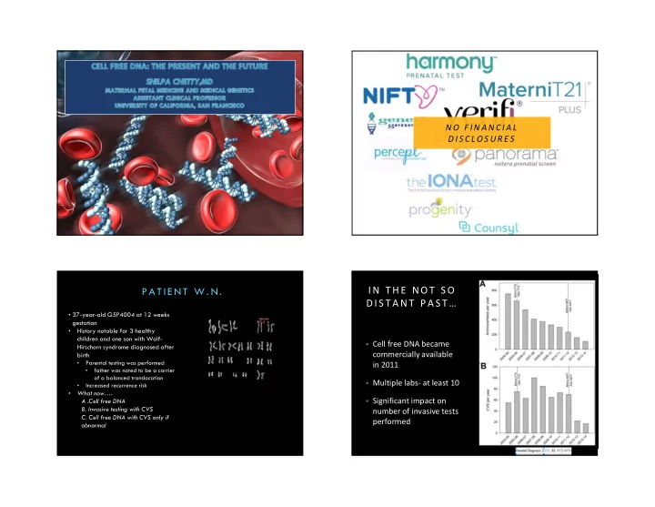
P A T I E N T W . N . IN T H E NO T S O D IS T A NT P A S T … • 37-year-old G5P4004 at 12 weeks gestation • History notable for 3 healthy children and one son with Wolf- • Cell free DNA became Hirschorn syndrome diagnosed after commercially available birth • Parental testing was performed in 2011 • father was noted to be a carrier of a balanced translocation • Multiple labs- at least 10 • Increased recurrence risk • What now…. • Significant impact on A .Cell free DNA B. Invasive testing with CVS number of invasive tests C. Cell free DNA with CVS only if performed abnormal 1
10/19/2017 C E L L F R E E D N A - A B I T O F B A C K G R O U N D • Results from apoptosis • Origin thought to be primarily placenta • Circulate in maternal plasma • Made up of short segments of fetal DNA (<200 base pairs) Cell Free DNA in Maternal Plasma: Characteristics cfDNA : Common Clinical Applications • Aneuploidy: detect abnormal ratio of a particular • cfDNA represents ~10-15% of total DNA in maternal plasma (Lo 1998, chromosome Chiu 2011) • Reliably detected after 7 wks gestation (Birch 2005) • Sex determination • Higher concentrations late in gestation • Short half life (16 min), undetectable by 2 hrs postpartum (Lo 1999) • Isoimmunization: noninvasively determine fetal Rh type 2
10/19/2017 cf DNA: The Future D I F F E R I N G T E C H N I Q UE S U S E D B Y D I F F E R E N T L A B S • Massively parallel shotgun sequencing (MPSS) • Microdeletions • Comprehensive • Easy to add additional analyses • Whole genome testing • Targeted sequencing • Less expensive • Single gene disorders • Single nucleotide polymorphisms (SNP) analysis • Can distinguish maternal from fetal DNA • Can identify triploidy, vanishing twin U N D E R S T A N D IN G T H E M E T H O D S F E T A L F R A C T I O N • Cell free DNA is isolated from maternal plasma • Total cfDNA is sequenced and The portion of millions of sequence reads fetal DNA in are generated maternal circulation • cfDNA • Sequence reads are aligned to the reference genome • Aligned reads are counted to determine chromosome number Prenatal Diagnosis Curr Genet Med Rep 2013 Jun; 1: (2):113-121 2012; 32(13) p 1233-41 3
10/19/2017 The less fetal DNA, the harder to tell normal from abnormal Cell free DNA screening: Biologic Challenges • False negatives: • False positives: • Low level of fetal • Unrecognized or “vanishing” DNA twin • Placental mosaicism • Placental mosaicism • Maternal genetic • Low level maternal variation (copy mosaicism, esp sex number variants) chromosomal • Maternal genetic variation (copy number variants) • Maternal malignancy C E L L F R E E D N A S C R E E N I N G : B I O L O G I C P E R F O R M A N C E O F C F D N A F O R O T H E R A N E U P L O ID I E S C H A L L E N G E S • Failed results: • Increased BMI (BMI >35) • Low level of fetal DNA • Fetal aneuploidy The false positives add up…. 4
10/19/2017 Microdeletions are More Common Than Down Syndrome for R A T E S O F A N O M A L I E S B Y M A T E R N A L A G E Women Under 40 Adapted from: Snijders, et al. Ultrasound Obstet Gynecol 1999;13:167–170 S H O U L D A L L W O M E N B E O F F E R E D M IC R OD E L E T IO N S A RE R A R E S C R E E NI N G F O R M I C R O D E L E T I O N S ? 5
10/19/2017 • Microdeletions can be detected by cfDNA, but… • Difficult to validate • Much of the data on in vitro samples • Clinical testing includes small number of syndromes • 22q, 1q36, 5p-, 4p-, 15q11-13 (Prader-Willi/Angelman ) Ultrasound Obstet Gynecol, 2016 Prevalence of many microdeletion syndromes is unknown • Many such clinical syndromes are caused by different molecular mechanisms • Only deletions detected • Smaller deletions more difficult to detect • 22q syndrome (~1/4000) • 85% have 3Mb deletion, 15% smaller • “97% detection” refers to only 3Mb deletion 6
10/19/2017 Sequenced 565 samples: 31 with CNV and 534 normal • Detected 83% with CNV >6Mb but only 20% (2/10) that were <6Mb • 2/534 false positives • More sequencing = higher sensitivity cfDNA for microdeletions is NOT a noninvasive T H E F A L S E P OS IT I V E S A D D U P … microarray Prevalence of 5 microdeletion syndromes: ~1/2500 Prevalence of CNV by microarray: 1/60 Detection rate of cfDNA for all pathogenic CNV: 4.2% 7
10/19/2017 T H E F U T U R E : N O N I N V A S I V E W H O L E G E N OM E S E Q U E N C I N G • Coding and non coding portions • Single nucleotide variants • Screening for these microdeletions has not been validated in clinical studies. • Deletions and Duplications • Routine cell-free DNA screening for microdeletion syndromes should not be performed. • Copy number variants 8
10/19/2017 I S M OR E A L W A Y S B E T T E R ? • Ehrich et al. (2017) reports on the clinical experience of one lab’s experience with • Obtain information about the the first 10,000 cases entire fetal genome • reports on copy number variations >7 Mb • Raises both practical and ethical • 25% of samples were sent due to an ultrasound abnormality issues • Screen-positive test results were reported in 554 cases, leading to a screen- • May give information regarding positive rate of approximately 5.4% (compared with 2.3% in traditional cfDNA screening) risk for adult onset conditions that are not relevant • However, pregnancy outcomes not available so no data on false positive or false negatives Non-Invasive Single Gene Tests • Maternal and fetal cell free DNA cannot be easily distinguished • mother and fetus may share the same mutation • Can identify de novo or paternal gene mutation circulating in maternal blood • An affected fetus will have an overrepresentation of the mutant allele in maternal • N=47 cases plasma DNA • Correct in 46 (96.2%) • Useful tool in 3 rd trimester to distinguish IUGR from achondroplasia • Ideal for autosomal dominant paternally inherited conditions • Useful if testing for an autosomal recessive condition and parents have different mutations 9
10/19/2017 C E L L F R E E D N A T E S T I N G F O R S I N G L E G E N E B A C K T O OU R C A S E … D I S O R D ER S • Our patient underwent cell free DNA testing with microdeletions • Current clinically available for limited conditions • Her results were normal so she declined any further testing • Achondroplasia • Level II ultrasound was notable for unilateral cleft lip and • Thanatophoric dysplasia palate and abnormal profile • Apert syndrome, CF • Amniocentesis confirmed deletion of 8.9 MB deletion on 4pter consistent with Wolf-Hirshorn Current guidelines? A C O G / S M F M 2 0 1 5 • Conventional screening is most appropriate first line screen for most patients • Ethically any patient may choose cfDNA screening, but should be counseled regarding limitations and benefits • Diagnostic testing is required to confirm abnormal results before irreversible decisions Special thanks to Meg Autry, MD & Mary Norton, MD • Testing for microdeletions and in twins should not be performed 10
10/19/2017 11
Recommend
More recommend