SLIDE 1 1
#1
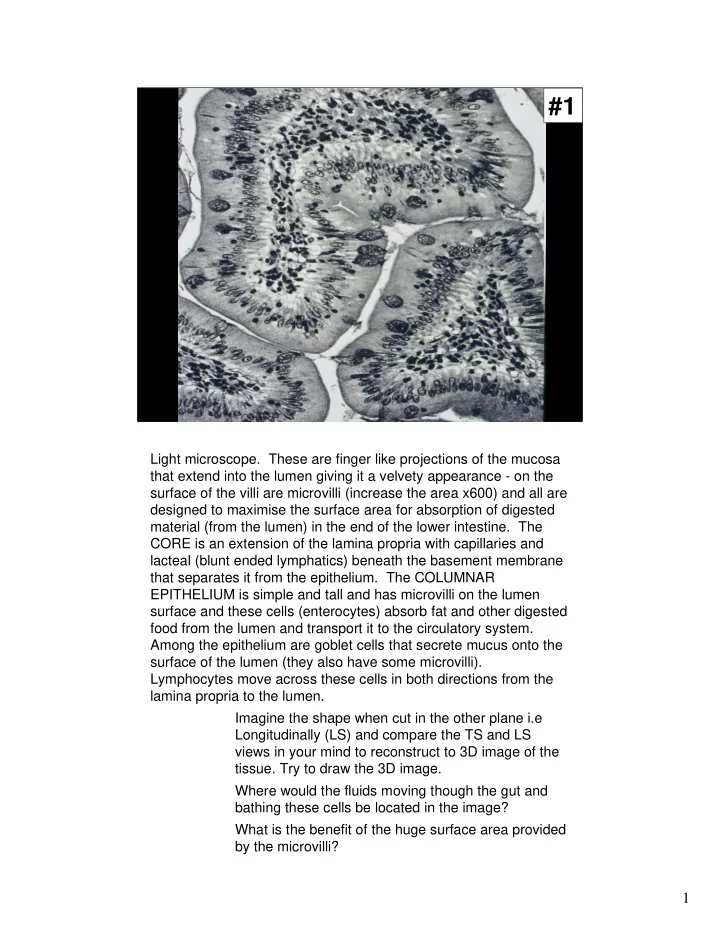
Light microscope. These are finger like projections of the mucosa that extend into the lumen giving it a velvety appearance - on the surface of the villi are microvilli (increase the area x600) and all are designed to maximise the surface area for absorption of digested material (from the lumen) in the end of the lower intestine. The CORE is an extension of the lamina propria with capillaries and lacteal (blunt ended lymphatics) beneath the basement membrane that separates it from the epithelium. The COLUMNAR EPITHELIUM is simple and tall and has microvilli on the lumen surface and these cells (enterocytes) absorb fat and other digested food from the lumen and transport it to the circulatory system. Among the epithelium are goblet cells that secrete mucus onto the surface of the lumen (they also have some microvilli). Lymphocytes move across these cells in both directions from the lamina propria to the lumen. Imagine the shape when cut in the other plane i.e Longitudinally (LS) and compare the TS and LS views in your mind to reconstruct to 3D image of the
- tissue. Try to draw the 3D image.
Where would the fluids moving though the gut and bathing these cells be located in the image? What is the benefit of the huge surface area provided by the microvilli?
SLIDE 2 2
#2
Similar view of ileum but stained with toluidine blue Is a plastic resin section. Magnification Think of a nucleus as about 5 µm (0.005mm) in
- diameter. In the photo they are about 2mm (2000µm). Therefore
divide 2000 by 5 = 400. i.e is magnified about 400 times. A= brush border/ microvilli, B=core of lamina propria with vessel which could be a lacteal or a blood vessel, C=goblet cell, D=lymphocyte. COMMENT ON FIXATION compare the histological detail with #1
SLIDE 3
3
#3
Electron micrograph of brush border. The apical (luminal) ends of 2 epithelial cells are shown with their microvilli on the surface, smooth endoplasmic reticulum in the cytoplasm and a desmosome junctional complex connecting the 2 cells. Magnification is about 50 x more than #12 (i.e width of brush border is about 50mm cf 1mm) therefore magnification is about 20,000. Note: really high power EM view (x 85,000) of microvilli on p457, R&R. WHERE LOCATED IN #1 & 2?
SLIDE 4
4
#4
PERIPHERAL NERVE. Cross section of myelinated and unmmyelinated nerves and support Schwann cells (p264 - 270, R&R; p112 G&H). Electron microscope (x50,000). A= axon with E = thick myelin sheath and C = peripheral cytoplasm of Schwann cell with basal lamina separating it from the supporting connective tissue (P267 R&R). B = smaller axons of unmyelinated nerves lying within grooves but enclosed by the Schwann cell cytoplasm. What cell type makes the myelin sheath?
SLIDE 5 5
#5
- LUNG. Electron micrograph of type II alveolar cell with alveolar
spaces and red blood cells. (p542-546 R&R). Magnification Estimation: nucleus measures about 30mm (=30,000 µm) therefore magnification is 6,000. Scale bar of 20mm represent 2µm, therefore magnification actually is 10,000. The type II alveolar cell (pneumocyte) has very distinctive multilamella bodies that make granules of phospholipid surfactant that are secreted into the alveolar air space, the cell has a dome shaped apical surface with some short microvilli. The surfactant covers the alveolar epithelium and reduces the surface tension and prevents the alveoli collapsing on exhalation. The alveolar epithelium is composed mainly of type I (95%) and type II (5%) alveolar cells. Connective tissue cells and several thin walled capillaries with RBC are present.
SLIDE 6 6
#6
- LIVER. Light microscope view of hepatocytes, sinusoids and
hepatic vein. Reticulin stain. (p498-504, p521, R&R; p232 G&H). Magnification x 100.
SLIDE 7 7
#7
THYROID FOLLICLE. Electron microscope view of colloid and cuboidal follicular cells (p603 - 606, p612, 630 R&R; p157 G&H). The follicular cells have lots of vesicles and ER. The colloid consists of thyroglobulin which is secreted by the follicular cells and is the inactive storage form of the hormones T4 and T3. Only after further cellular processing are the thyroid hormones liberated from the colloid and passed out to the capillaries. Apical pseudopods within the colloid show that the follicular cells are removing the
- colloid. The thyroid is unique amongst endocrine glands because it
stores its secretory product extracellularly. Magnification Estimate gives about 8,000, scale bar gives 35,000 divided by 5 = 7,000.
SLIDE 8
8
#8
BLOOD CELLS. Light microscope of blood smears stained with Wright’s stain (p188 - 207, p210-213 R&R; p71-73 G&H). A = Neutrophil (polymorphonuclear leukocyte - lobulated nuclei), B = Eosinophil (pink granules and sausage-shaped nucleus), C = RBC (erythrocyte - many of these, no nucleus, clear area is thinnest area of biconcave disk, long diameter is 7-8), D = Monocyte (large cell, acentric kidney shaped nucleus and lack of granules) , E = Platelet (tiny bits of cytoplasm 2µm in diameter, no nucleus). x 1,000.
SLIDE 9 9
#9
#9 & #10
- CARTILAGE. Longitudinal section through the trachea showing
hyaline cartilage (blue) and the dense connective tissue of the perichondrium (pink) surrounded by epithelium (brown) (p132 - 138, p140-149, 177, R&R; 53, 61 G&H). #8 Low power (x 100) stained with Alcian blue, light microscope. Shows chondrocytes and matrix
- proteins. Note the pronounced lacunae around the chondrocytes
due to the processing. In contrast in #10 in the EM (x 10,000) the lacuna is less pronounced and the chondrocytes are closely associated with the matrix. SEE EM PICTURE ON GROUNDS’ FLOOR (AT FOOT OF STAIRS) = PICTURE #33
SLIDE 10 10
#10
#9 & #10
- CARTILAGE. Longitudinal section through the trachea showing
hyaline cartilage (blue) and the dense connective tissue of the perichondrium (pink) surrounded by epithelium (brown) (p132-138, p140-149, 177, R&R; 53, 61 G&H). #8 Low power (x 100) stained with Alcian blue, light microscope. Shows chondrocytes and matrix
- proteins. Note the pronounced lacunae around the chondrocytes
due to the processing. In contrast in #10 in the EM (x 10,000) the lacuna is less pronounced and the chondrocytes are closely associated with the matrix. SEE EM PICTURE ON GROUNDS’ FLOOR (AT FOOT OF STAIRS) = PICTURE #33
SLIDE 11 11
#11
- BONE. Undecalcified ground compact bone, paraffin cross section
treated with Indian ink to show the features x 100. ( p150-169, 171 R&R; p56-59 G&H). Osteocytes in lacunae with canaliculi, the lamelli of deposited bone matrix and Haversian canals are shown.
SLIDE 12
12
#12
COLLAGEN FIBRILS. Very high power EM showing the typical 64nm banding of collagen. (p96-104, p98, R&R) Magnification 100nm = 0.1µm measures 20,000µm (20 mm), therefore = 200,000.
SLIDE 13
13
#13
CILIATED EPITHELIUM on microvilli . There are no cilia on the epithelium in the gut but there are on the vas deferens and the fallopian tube and in the respiratory tracts (see p537 R&R). Scanning electron micrograph (x 400). Note the long cilia on some cells (all waving in the same direction at the time of fixation) whereas only short microvilli are present on other cells.
SLIDE 14 14
#14
JUNCTION OF STRATIFIED SQUAMOUS EPITHELIUM AND COLUMNAR EPITHELIUM. Light microscope stained with Alcian blue and haematoxylin (x 400). Beneath the epithelium is loose connective tissue and glands. No
- cilia. Probably the cervix (Fig 4 p727 R&R) at the junction of the
vagina (squamous) and the cervical end of the uterus (columnar - p723, R&R) (p267 - 271, G&H)