Voluntary Movement II. 1. Primary motor cortex: how are movement - - PDF document

voluntary movement ii
SMART_READER_LITE
LIVE PREVIEW

Voluntary Movement II. 1. Primary motor cortex: how are movement - - PDF document

Voluntary Movement II. 1. Primary motor cortex: how are movement parameters coded Cortical representation of Distal movements CM neurons. movements and Population coding. 2. Premotor areas higher order features of parameters.


slide-1
SLIDE 1

1

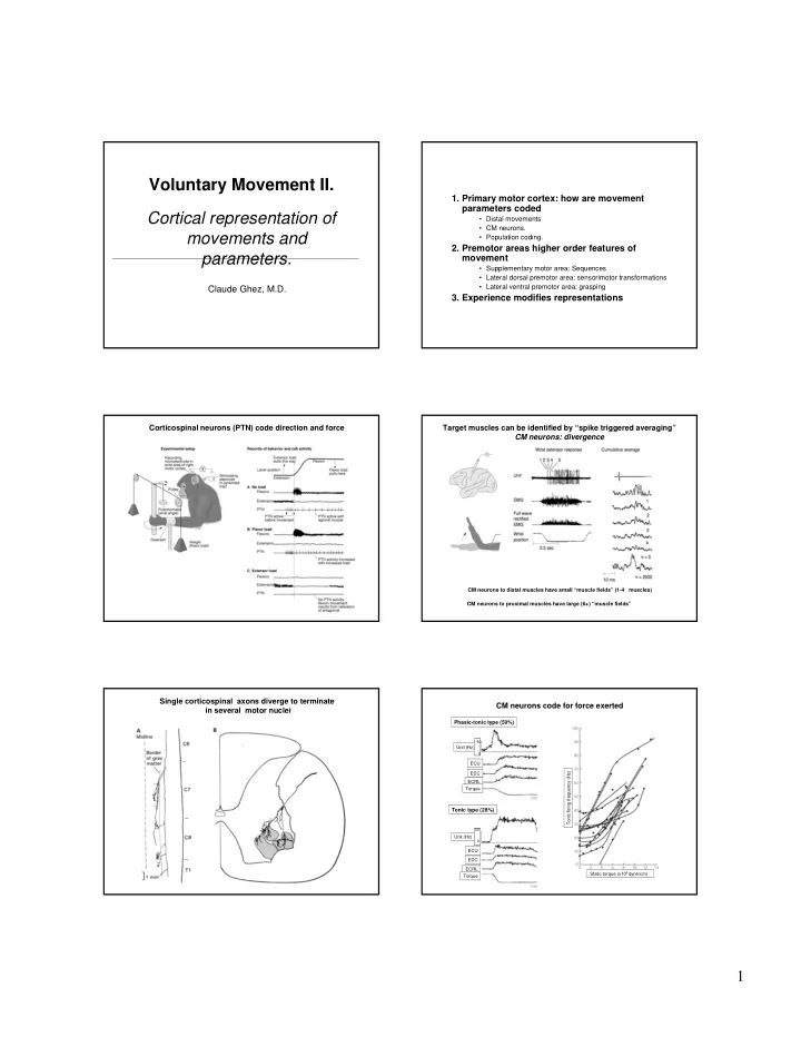
Voluntary Movement II. Cortical representation of movements and parameters.

Claude Ghez, M.D.

  • 1. Primary motor cortex: how are movement

parameters coded

  • Distal movements
  • CM neurons.
  • Population coding.
  • 2. Premotor areas higher order features of

movement

  • Supplementary motor area: Sequences
  • Lateral dorsal premotor area: sensorimotor transformations
  • Lateral ventral premotor area: grasping
  • 3. Experience modifies representations

Corticospinal neurons (PTN) code direction and force Target muscles can be identified by “spike triggered averaging” CM neurons: divergence

CM neurons to distal muscles have small “muscle fields” (1-4 muscles) CM neurons to proximal muscles have large (6+) “muscle fields”

Single corticospinal axons diverge to terminate in several motor nuclei

Static torque (x105 dyne/cm) Tonic firing frequency (Hz)

Tonic type (28%)

Unit (Hz) ECU EDC ECRL Torque Unit (Hz)

50 50

ECU EDC ECRL Torque

Phasic-tonic type (59%)

CM neurons code for force exerted

slide-2
SLIDE 2

2

CM neuron EMG of muscle

Precision grip Power grip

CM neurons are preferentially recruited for tasks requiring topographical precision

Intact (normal) After section of corticospinal fibers

Section of pyramidal tracts in monkeys produces loss of independent “individuated” digit control Neurons in proximal motor cortex regions are broadly tuned for direction Movement direction can be coded precisely by the population responses of broadly tuned neurons Primary motor cortex receives direct input from 5 premotor areas These premotor areas also project to the spinal cord “Self initiated”voluntary movement are preceded by premotor activation: early evidence from evoked potentials

slide-3
SLIDE 3

3

Mental rehearsal of finger sequence Motor cortex Supplementary motor area Sensory cortex Repeated simple finger flexion Repeating sequence finger-thumb apposition

Planning movement sequence without moving activates SMA First neuroimaging data

Primary motor cortex Lateral premotor area Supplementary motor area

1st key touch 1st key touch 1st key touch Visual Cue Learned Sequence

Activation of motor areas depend different on behavioral context Supplementary motor area neurons code movements in specific context of movement sequence.

Cell fires with pull followed by turn but not followed by pull Cell fires with turn followed by pull and push but not just with pull

Reaching Grasping

Primary Motor

Separate pathways convey visual inputs to premotor areas for reaching and grasping

Instruction: Left Instruction: Right LED= trigger signal Panel= instruction signal Instruction stimulus Trigger stimulus Instruction stimulus Trigger stimulus

Instructed delay task: coding of “preparatory set” for directed reach by dorso-lateral premotor neurons

Contralateral movement Ipsilateral movement

  • Rec. Ventral PM

Precision grip Power grip

Neurons in ventral premotor area (PMv) code for hand configuration of grasp

slide-4
SLIDE 4

4

“Mirror neurons” in PMv represent types of movement independent of its actualization: motor vocabulary Practice and learning of finger sequence can alter motor representations in primary motor cortex

Elbow & shoulder

Infarct Infarct

Damage to local region of motor cortex induces change in representation of nearby areas

Preinfarct Post infarct with Rehabilitative therapy

Digit Wrist & forearm Digit, wrist & forearm Proximal No response

Infarct Infarct

Motor practice can alter functionality and motor mapping In motor cortex