visual system in ms
play

Visual system in MS Optic neuritis (ON): Inflammatory - PDF document

6/9/2014 Multifocal Visual Evoked Potentials (mfVEP) and Ganglion Cell Inner Plexiform Thickness (GCIPT) in Relapsing Remitting Multiple Sclerosis (RRMS) Divya Narayanan, Han Cheng, Rosa Tang, Laura Frishman University of Houston Houston, TX


  1. 6/9/2014 Multifocal Visual Evoked Potentials (mfVEP) and Ganglion Cell Inner Plexiform Thickness (GCIPT) in Relapsing Remitting Multiple Sclerosis (RRMS) Divya Narayanan, Han Cheng, Rosa Tang, Laura Frishman University of Houston Houston, TX Visual system in MS • 
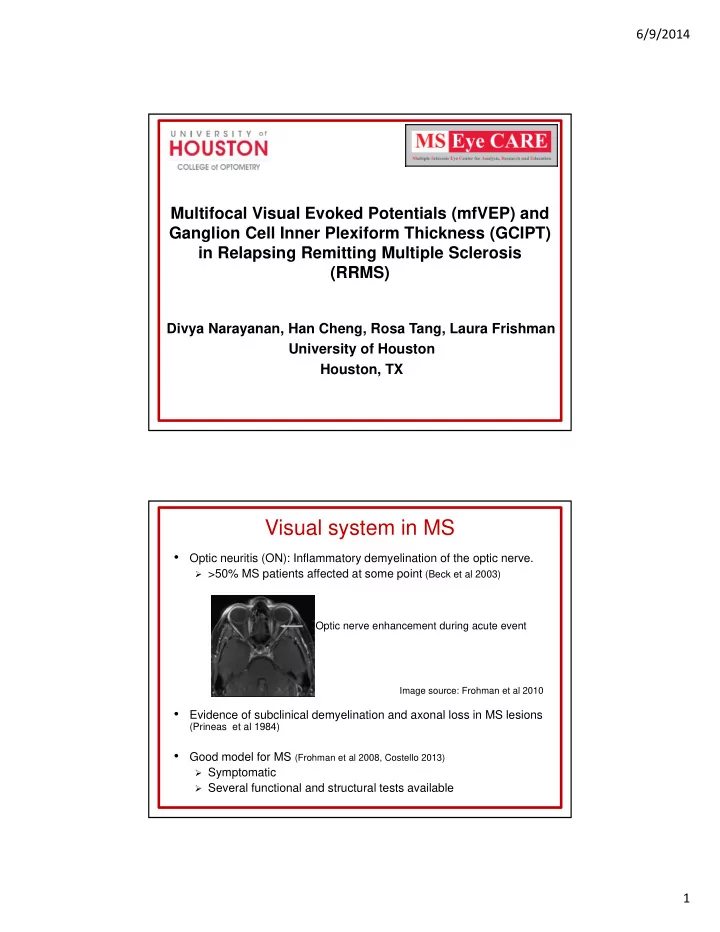
Optic neuritis (ON): Inflammatory demyelination of the optic nerve.  >50% MS patients affected at some point (Beck et al 2003) Optic nerve enhancement during acute event Image source: Frohman et al 2010 • Evidence of subclinical demyelination and axonal loss in MS lesions (Prineas et al 1984) • Good model for MS (Frohman et al 2008, Costello 2013)  Symptomatic  Several functional and structural tests available 1

  2. 6/9/2014 Clinical tests to assess visual system Functional tests Structural tests • Optical coherence tomography Subjective (OCT) • Contrast sensitivity (CS) • Humphrey visual fields (HVF) Objective • Traditional visual evoked potential (tVEP) • Multifocal visual evoked potential (mfVEP) Contrast sensitivity Humphrey visual fields MD -5.13 P < 1% • Pelli-Robson contrast measured from 0 to 2.25 log • HVF 30-2 or 24-2 were units in 0.15 log unit steps performed. Image source: http://www.psych.nyu.edu/pelli/pellirobson 2

  3. 6/9/2014 Visual evoked potential (VEP) • Non-invasive measure of electrical responses generated by visual cortex • Amplitude : Loss of nerve fibers reduces amplitude • Latency : Demyelination delays signals Image source: Sensory testing systems Traditional VEP Pattern-reversal stimulus Response • 15’, 60’ and 120’ check sizes • P100 amplitude and latency • 2 reversals per second measured • Provides summed responses dominated from macular region 3

  4. 6/9/2014 Multifocal VEP (MfVEP) Stimulus Response • 60-sectors,scaled for cortical • Amplitude: Log signal-to-noise magnification ratio (logSNR) • Multiple VEPs simultaneously • Relative latency: Cross-correlation recorded from 60 local regions of subject’s waveform and normative template Hood et al 2004 MfVEP probability plots • Responses from each sector are compared to the norms and marked as normal or abnormal • Topographic read out of the extent of damage AMP LAT Saturated: p<0.01 Desaturated: p<0.05 Red: Left eye abnormal Blue: Right eye abnormal 4

  5. 6/9/2014 SD-OCT Peripapillary retinal nerve fiber layer thickness (RNFLT) and macular ganglion cell inner plexiform layer thickness (GCIPT) measures were obtained GCIP Image Source Leung et al 2013 Syc et al 2011 Purpose To compare various functional and structural measures in RRMS eyes, especially those without a history of ON 5

  6. 6/9/2014 Methods • 90 RRMS patients had CS, HVF, mfVEP, OCT • Mean age: 40.8 ± 10.5 years • Mean MS duration: 6.5 ± 7.4 years  58 ON eyes (last ON>6months)  Time since last ON: 3.8 ± 5.0 years  105 non-ON eyes • 30 patients (19 ON, 30 non-ON eyes) also had tVEP • 40 age-matched normal controls Analysis Criteria for classifying MS eyes as abnormal • CS , MfVEP and tVEP classified as abnormal if <5% of norms • HVF (MD), GCIPT and RNFLT classified as abnormal if <5% of machine norms 6

  7. 6/9/2014 Results ON : Percent of abnormal eyes detected by functional tests ** p<0.01 7

  8. 6/9/2014 ON : GCIPT detected more abnormal eyes than RNFLT S T N I Macula Optic disc Macula Optic disc * p<0.05 Non-ON : MfVEP detected more abnormal eyes than TVEP ** p<0.01 8

  9. 6/9/2014 Non-ON : GCIPT detected more abnormal eyes than RNFLT S T N I Optic disc Macula * p<0.05 Among non-ON eyes with abnormal mfVEP LAT, 65% had delay in the central region that corresponds to GCIPT 65% MfVEP region GCIPT region 7.3° horizontal 6.0° vertical* 8° horizontal 6.7° vertical *Scaled for retinal ganglion cell displacement (Drasdo et al 2007) 9

  10. 6/9/2014 Correlation between functional tests vs GCIPT MfVEP: Correlated with GCIPT in both ON and non-ON TVEP: Correlated with GCIPT in ON but not in non-ON 10

  11. 6/9/2014 CS: Correlated with GCIPT in both ON and non-ON HVF: Correlated with GCIPT in ON but not in non-ON Conclusion • MfVEP detected more abnormal eyes than other functional tests (3 times more than tVEP in non-ON) • GCIPT detected more abnormal eyes than ARNFLT and TRNFLT • Pelli-Robson CS and mfVEP are more reflective of the structural alterations than HVF and tVEP, especially in non- ON eyes • MfVEP and GCIPT offer complementary information on the integrity of the visual pathway and are useful for detecting subclinical neuronal defects in MS 11

  12. 6/9/2014 Acknowledgement • Organizers: Whitaker Research • Dr Han Cheng • Dr Laura Frishman Track • Dr Rosa Tang • CMSC scholarship • Dr Ronald Harwerth • NIH P30 EY 007551 • Courtney Perry • Bobby Saenz • NIH grant T35 EY 007088 • Fight for Sight fellowship • MS Eye CARE team • Minnie Flaura Turner memorial fund for impaired vision research 12

Download Presentation
Download Policy: The content available on the website is offered to you 'AS IS' for your personal information and use only. It cannot be commercialized, licensed, or distributed on other websites without prior consent from the author. To download a presentation, simply click this link. If you encounter any difficulties during the download process, it's possible that the publisher has removed the file from their server.

Recommend


More recommend