
SLIDE 1
1
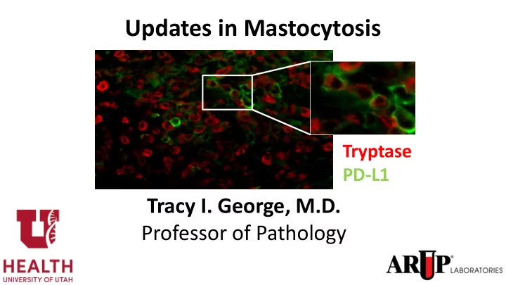
Updates in Mastocytosis Tryptase PD-L1 Tracy I. George, M.D. - - PowerPoint PPT Presentation
Updates in Mastocytosis Tryptase PD-L1 Tracy I. George, M.D. Professor of Pathology 1 Disclosure: Tracy George, M.D. Research Support / Grants None Stock/Equity (any amount) None Consulting Blueprint Medicines Novartis Employment ARUP
1
Research Support / Grants None Stock/Equity (any amount) None Consulting Blueprint Medicines Novartis Employment ARUP Laboratories Speakers Bureau / Honoraria None Other None
Polycythemia Vera Essential Thrombocythemia Primary Myelofibrosis Chronic Neutrophilic Leukemia Chronic Eosinophilic Leukemia, NOS Hypereosinophilic Syndrome Mast Cell Disease MPNs, unclassifiable Chronic Myelomonocytic Leukemia Atypical Chronic Myeloid Leukemia Juvenile Myelomonocytic Leukemia MDS/MPN, unclassifiable Chronic Myelogenous Leukemia Myeloid neoplasms associated with PDGFRA rearrangement Myeloid neoplasms associated with PDGFRB rearrangement Myeloid neoplasms associated with FGFR1 rearrangement (EMS)
Acute Myeloid Leukemia Myelodysplastic Syndromes Myeloproliferative Neoplasms MDS/MPN Myeloid or lymphoid neoplasms associated with eosinophilia and abnormalities of PDGFRA, PDGFRB, or FGFR1
Polycythemia Vera Essential Thrombocythemia Primary Myelofibrosis Chronic Neutrophilic Leukemia Chronic Eosinophilic Leukemia, NOS MPN, unclassifiable Chronic Myelomonocytic Leukemia Atypical Chronic Myeloid Leukemia Juvenile Myelomonocytic Leukemia
MDS/MPN with ring sideroblasts and thrombocytosis
MDS/MPN, unclassifiable Chronic Myeloid Leukemia Myeloid/lymphoid neoplasms with PDGFRA rearrangement Myeloid/lymphoid neoplasms with PDGFRB rearrangement Myeloid/lymphoid neoplasms with FGFR1 rearrangement Myeloid/lymphoid neoplasms with PCM1-JAK2
Acute Myeloid Leukemia Myelodysplastic Syndromes Myeloproliferative Neoplasms MDS/MPN Myeloid/ lymphoid neoplasms with eosinophilia and gene rearrangement Mastocytosis
Morgado JM, Sánchez-Muñoz L, Teodósio CG, Jara-Acevedo M, Alvarez-Twose I, Matito A, Fernández-Nuñez E, García-Montero A, Orfao A, Escribano L. Immunophenotyping in systemic mastocytosis diagnosis: 'CD25 positive' alone is more informative than the 'CD25 and/or CD2' WHO criterion. Mod Pathol. 2012;25:516-21.
Morgado JM, Sánchez-Muñoz L, Teodósio CG, Jara-Acevedo M, Alvarez-Twose I, Matito A, Fernández-Nuñez E, García-Montero A, Orfao A, Escribano L. Immunophenotyping in systemic mastocytosis diagnosis: 'CD25 positive' alone is more informative than the 'CD25 and/or CD2' WHO criterion. Mod Pathol. 2012;25:516-21.
Horny H-P et al. Mastocytosis. In: Swerdlow SH et al (eds). WHO Classification of Tumours of Haematopoietic and Lymphoid Tissues. Revised 4th Edition. IARC Press, Lyon, 2017
– Indolent systemic mastocytosis – Smoldering systemic mastocytosis – Systemic mastocytosis with associated hematologic neoplasm – Aggressive systemic mastocytosis – Mast cell leukemia
P Valent et al. Refined diagnostic criteria and classification of mast cell leukemia and myelomastocytic leukemia: a consensus proposal. Ann Oncol 2014;24(9):1691-1700.
?Diagnosis
Cutaneous mastocytosis only
Work up:
Berezowska S, Flaig MJ, Rueff F, et al. Adult-onset mastocytosis in the skin is highly suggestive of systemic mastocytosis. Mod Pathol 2014;27:19.
Theoharides TC et al. N Engl J Med 2015;373:163-172.
Clinically Relevant Mediators Released from Mast Cells and Putative Effects.
17
Longly Jr BJ, et al. PNAS. 1999;96:1609-1614; Nakagomi N, et al. Lab Invest. 2007;87:365-371; Horny HP, et al. Pathobiology. 2007;74:121-132; Garcia-Montero AC, et al. Blood. 2006;108:2366-2372.
KIT kit kit kit kit
ASM (1+ “C”=cytoreductive requiring findings)
function, ascites, +/- portal hypertension
lesions, and/or pathological fractures
infiltrates, hypoalbuminemia
Mast cell leukemia ≥20% mast cells
SM + AHN
associated hematological neoplasm
George and Horny. Systemic mastocytosis. Hematol Oncol N Am 25 (2011): 1067.
Normal/reactive/well-differentiated Atypical type I Atypical type II Metachromatic blast
George and Horny. Systemic mastocytosis. Hematol Oncol N Am 25 (2011): 1067.
Normal/reactive/well-differentiated Atypical type I Atypical type II Metachromatic blast
tryptase
CD117 ASM- MDS/MPN,U tryptase ASM-CMML
Bone marrow aspirate
Laboratory values: Hb: 8.8 g/dL WBC, PLT: Normal Serum tryptase: 763
Blood smear
CD117 CD25 CD25 CD2 Tryptase IHC positive KIT D816V positive
age and sex-matched US population’s survival for the entire cohort
29
Lim, KH. Blood. 2009 Jun 4;113(23):5727.
Years From Diagnosis
ISM (n = 159) 100 80 60 40 20 10 20 30
Survival
ASM (n = 41) AHNMD (n = 138) MCL (n = 4) Matched Controls
Juxtamembrane domain Transmembrane domain Extracellular ligand-binding domain Tyrosine kinase domain 1 Tyrosine kinase domain 2 Kinase insert
NH2 COOH
Dimerization domain GIST Exon 9
GIST: Gastrointestinal stromal tumors; SM: Systemic Mastocytosis; AML: acute myelogenous leukemia; NK/T-CL: Natural killer/T-cell lymphoma
AML (Asp 419) Exon 8 SM V560G; GIST Exon 11 Sinonasal NK/T-CL V559I,E561K GIST Exon 13 SM D816V,Y; GIST Exon 17 AML: D816V,Y Germ cell tumors: D816H Sinonasal NK/T-CL D816N, D825A
D816V~80% Rare Imatinib sensitive Imatinib resistant
– Observation – Topical therapies for cutaneous disease – Symptomatic noncytoreductive therapies – Cytoreductive therapy
31
Akin C, et al. Exp Hematol. 2003;31:686-692; Douglass JA, et al. Allergy. 2010; 65:924-932; Pardanani A, et al. Curr Opin Hematology. 2010;17:125-132; Imatinib package insert.
all c-KIT-transformed Ba/F3 cell lines
due to expression of c-KIT D816V are inhibited by midostaurin
additional cell lines
32
Growney JD, et al. Blood. 2005;106:721-724; Gotlib J, et al. Blood. 2005;106:2865-2870.
IC50 for midostaurin: 44 nM IC50 for imatinib: > 1 uM
3H-Thymidine Incorporation ( %)
33
type c-KIT
cultured cord blood cell-derived mast cells
Growney JD, et al. Blood. 2005;106:721-724. Krauth M-T, et al. Clin Exp Allergy. 2009; 39:1711-1720].
June 30, 2016;374:2530-2541.
Gotlib J et al. N Engl J Med 2016;374:2530-2541.
C
Gotlib J et al. N Engl J Med 2016;374:2530-2541.
Bone marrow mast cell burden Serum tryptase Spleen volume
Alk Phos Eos Monos
DeAngelo DJ, George TI, Linder A, Langford C, Perkins C, Ma J, Westervelt P, Merker JD, Berube C, Coutre S, Liedtke M, Medeiros B, Sternberg D, Dutriex C, Ruffie PA, Corless C, Graubert TJ, Gotlib J (2018). Leukemia, 32(2), 470-478.
CD25 expression on neoplastic mast cells by flow cytometry in patients #1, #2, and #3 before (A- C) and on day 28 (patient #1, D), day 89 (patient #2, E), and day 336 (patient #3, F) of midostaurin therapy.
A B D E C F
Patient #1 Patient #2 Patient #3
Midostaurin therapy is associated with sustained decreases in CD25 expression
CD25 expression on neoplastic mast cells by mean fluorescence intensity (MFI; A, C, E) and percent of mast cells positive for CD25 (B, D, F) over time in patient #1 (A-B), patient #2 (C-D), and patient #3 (E-F). The shaded area indicates time on midostaurin therapy.
20 40 60 80 100
40 90 140 190 240 290 340 390
%CD25 positive mast cells Days of treatment
1.E+03 1.E+04 1.E+05
10 30 50 70 90
CD25 MFI Days of treatment
50 100
10 30 50 70 90
% CD25 positive mast cells Days of treatment
1.E+02 1.E+03 1.E+04 1.E+05
40 90 140 190 240 290 340 390
CD25 MFI Days of treatment
1.E+03 1.E+04 1.E+05
50 100 150 200 250 300 350 400 450 500 550
CD25 MFI Days of treatment
20 40 60
50 100 150 200 250 300 350 400 450 500 550
% CD25 positive mast cells Days of treatment
A C B E F D
#1 #2 #3
Mutant Kit
Constitutive signaling
STAT5 pSTAT5
Transcription
CD25 Mutant Kit Other RTKs? STAT5 PKC412 pSTAT5 Other RTKs?
Changes in the intracellular localization of STAT5 with midostaurin
JM Morgado, O Perbellini,RC Johnson, C Teodosio, A Matito, I Alvarez-Twose, P Bonadonna, A Zamo M Jara-Acevedo, A Mayado, A Garcia-Montero, M Mollejo, TI George, R Zanotti, A Orfao, L Escribano, L Sanchez-Munoz. CD30 expression by bone marrow mast cells from different diagnostic variants of systemic mastocytosis. Histopathology 2013;63(6):780-7.
JM Morgado, O Perbellini,RC Johnson, C Teodosio, A Matito, I Alvarez-Twose, P Bonadonna, A Zamo M Jara-Acevedo, A Mayado, A Garcia-Montero, M Mollejo, TI George, R Zanotti, A Orfao, L Escribano, L Sanchez-Munoz. CD30 expression by bone marrow mast cells from different diagnostic variants of systemic mastocytosis. Histopathology 2013;63(6):780-7.
10 patients:
study
Baird JH, Verstovsek S, George TI, Reyes I, Abuel J, Perkins C, Langford C, Schroeder K, Gotlib J. Phase 2 study of Brentuximab Vedotin in patients with advanced systemic
Hatch EW, Geeze MB, Martin C, Salama ME, Hartmann K, Eisenwort G, Blatt K, Valent P, Gotlib J, Lee JH, Chen L, Ward HH, Lidke DS, George TI (2018). Variability of PD-L1 expression in
M Geeze, E Hatch, C Martin, S Perkins, K Hartmann, P Valent, J Gotlib, D Lidke. USCAP 2016 PD-1 PD-L1 Smoldering systemic mastocytosis PD-L1 PD-1 Mast cell leukemia
Indolent SM Indolent SM Indolent SM Mast cell leukemia Mast cell leukemia Aggressive SM
Mast cell leukemia, spleen Tryptase (red), PDL1 (green)
Cutaneous mastocytosis Mast cell leukemia, spleen Tryptase (red) PDL1 (green)
Diagnosis PD-L1 expression PD-1 expression SM 17/22 (77%) 0/25 CM 23/25 (92%) 4/27 (15%) MML 1/2 0/2 MMAS 0/3 0/3 MPN 0/16 0/17 MDS 0/18 0/18 MDS/MPN 0/5 0/5 Healthy/ reactive BM 0/15 0/21
Diagnosis PD-L1 expression PD-1 expression SM 17/22 (77%) 0/25 CM 23/25 (92%) 4/27 (15%) MML 1/2 0/2 MMAS 0/3 0/3 MPN 0/16 0/17 MDS 0/18 0/18 MDS/MPN 0/5 0/5 Healthy/ reactive BM 0/15 0/21 MCL 3/3 (100%) ASM 2/2 (100%) SM-AHN 9/12 (75%) SSM 1/2 (50%) ISM 3/4 (75%)
Jason Gotlib Dan Arber Susan Atwater Bruno Medeiros Athena Cherry
Chris Corless Ilana Kepten Jeffrey Tyner Peter Valent Hans-Peter Horny Karl Sotlar
Eric Hsi Timothy Graubert Dan Deangelo Natasha Savage Farrukh Awan REMA (Spanish Network on Mastocytosis)