
SLIDE 1
8/25/2017 1
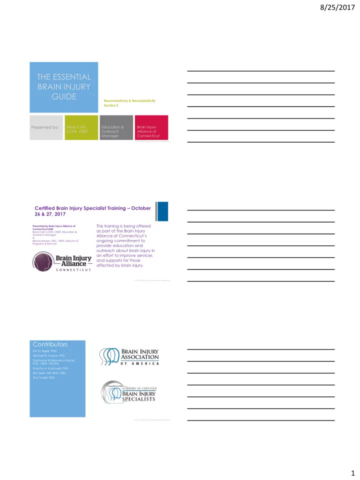
Neuroanatomy & Neuroplasticity Section 2
THE ESSENTIAL BRAIN INJURY GUIDE
Presented by:
Rene Carfi, LCSW, CBIST Education & Outreach Manager Brain Injury Alliance of Connecticut
Certified Brain Injury Specialist Training – October 26 & 27, 2017
This training is being offered as part of the Brain Injury Alliance of Connecticut’s
- ngoing commitment to
provide education and
- utreach about brain injury in
an effort to improve services and supports for those affected by brain injury.
Presented by Brain Injury Alliance of Connecticut staff: Rene Carfi, LCSW, CBIST, Education & Outreach Manager & Bonnie Meyers, CRC, CBIST, Director of Programs & Services
Contributors
Erin D. Bigler, PhD Michael R. Hoane, PhD Stephanie Kolakowsky-Hayner, PhD, CBIST, FACRM Dorothy A. Kozlowski, PhD Eric Spier, MD, BIM, CBIS Tina Trudel, PhD