SLIDE 1 Th The Car ardiovascu cular ar Sy System: m: Th The Hea Heart
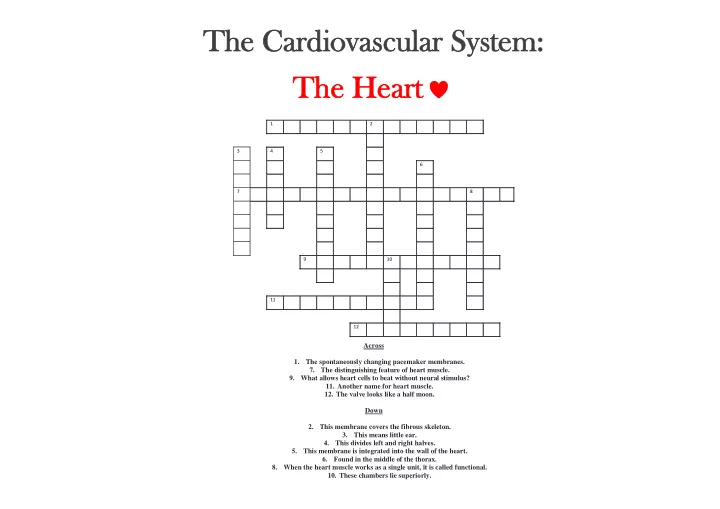
Across 1. The spontaneously changing pacemaker membranes. 7. The distinguishing feature of heart muscle. 9. What allows heart cells to beat without neural stimulus?
- 11. Another name for heart muscle.
- 12. The valve looks like a half moon.
Down 2. This membrane covers the fibrous skeleton. 3. This means little ear. 4. This divides left and right halves. 5. This membrane is integrated into the wall of the heart. 6. Found in the middle of the thorax. 8. When the heart muscle works as a single unit, it is called functional.
- 10. These chambers lie superiorly.
1 2 3 4 5 6 7 8 9 10 11 12
SLIDE 2 The Cardiovascu scular System em: The Heart
1
P R E P O T
2
E N T I A L S N
3 A
4
S
5
E D
U
E P O
6
M
R
P I C E
7 I N
T E R C A L A T E D D I
8
S C S
C
U A R I Y
L
M R D A N
E
D I S C
S
I U T Y
9
A U T O M
10
A T I C I T Y M T N I R U U
11
M Y O C A R D I U M M U
12
S E M I L U N A R
SLIDE 3
CARDIOVASCULAR
Blood Vessels
SLIDE 4 What are the three major types of blood vessels?
- A. Aorta, Common Carotid Artery, Superior
Vena Cava
- B. Brachiocephalic Artery, Right Coronary Sinus,
Intraventricular Artery
- C. Arteries, Capillaries, Veins
- D. Arterioles, Venules, Veins
SLIDE 5 On the border between the Tunica media and the Tunica Externa, there are small blood vessels supplying O2 and nutrients to the wall of the artery. What are these called?
- A. Lumen
- B. Veins
- C. Vasa Vasorum
- D. Vasa Viserous
SLIDE 6
How many circulatory pathways are there? What are they?
SLIDE 7 Pulmonary Circulation
The pulmonary circulation functions only to bring into close contact with the (air sacs) of the lungs so that can be exchanged.
- A. O2, bronchi, nutrients
- B. Bronchi, O2, gases
- C. O2, alveoli, gases
SLIDE 8 Systemic Circulation
The systemic circulation provides the functional blood supply to all body tissues; that is, it delivers , , and other needed substances while carrying away and
SLIDE 9 All are deep while are both deep and superficial.
- A. Veins, Arteries
- B. Arteries, Veins
SLIDE 10 All blood vessels except capillaries have three layers. Capillaries are composed of the tunica
- nly.
- A. Externa
- B. Intima
- C. Media
SLIDE 11 What type of blood vessel can serve as “a blood reservoir”?
SLIDE 12 Vessels returning blood to the heart are:
- A. Superior and inferior vena cava
- B. Left pulmonary arteries
- C. Right pulmonary veins
- D. Right and left pulmonary veins
SLIDE 13 What type of capillary is A?
A. Fenestrated B. Sinusoidal C. Continuous
SLIDE 14 What type of capillary is C?
A. Continuous B. Sinusoidal C. Fenestrated
SLIDE 15 What type of capillary is B?
A. Fenestrated B. Continuous C. Sinusoidal
SLIDE 16 What do Pericytes do?
- A. Live off the host
- B. Maintain capillary
- C. Assist with constriction
SLIDE 17
What is another name for Pericyte?
SLIDE 18 What is the formula for Mean Blood Pressure?
- A. (SR•HR)/1000
- B. DBP+1/3PP
- C. CO/HR
- D. 220-Age
SLIDE 19 How much blood volume is in the veins?
- A. 80%
- B. 75%
- C. 60%
- D. 20%
SLIDE 20 Fenestrated Capillaries can be found in several places. Which of them are shown below?
- A. Liver
- B. Small Intestine
- C. Skin
- D. Bone Marrow
SLIDE 21 What causes Precapillary Sphincters to
- pen?
- A. Hypotension
- B. Edema
- C. Increased Blood Pressure
- D. Cardiomegaly
SLIDE 22 How many locations are used for Palpating Pulse?
SLIDE 23 What chemicals increase BP?
- A. Nitric Oxide
- B. Alcohol
- C. Norepinephrine and Epinephrine
- D. Histamine, prostacyclin, and kinins
SLIDE 24
Three types of Shock that were discussed in class:
SLIDE 25
What is this a picture of?
SLIDE 26
Name 3 Vasodilators
SLIDE 27 Systemic pressure in the right atrium?
- A. 5 mmHg
- B. 0 mmHg
- C. 100 mmHg
- D. 90 mmHg
SLIDE 28 Which of the following is/are a type of blood vessel?
- A. Arteries, Capillaries, Veins
- B. Lymphatic
- C. All of the above
SLIDE 29
What is this picture of?
SLIDE 30
What blood vessel is most abundant?
SLIDE 31
Lymphatics are found in all tissues except:
SLIDE 32
What are the 2 most important functions of the lymphatic system?
SLIDE 33
The thoracic duct arises from the cisterna chyli and drains _______.
SLIDE 34 Pathway of Blood
valve right ventricle
- Right ventricle pulmonary semilunar valve
arteries lungs
- Lungs pulmonary veins left
- Left
valve left ventricle
semilunar valves aorta
circulation BONUS
SLIDE 35
What is the physiological process involving the growth of new blood vessels from pre-existing vessels? BONUS
SLIDE 36 Fill in the blanks:
A. B. C.
BONUS
SLIDE 37 Question and Answer Samples and Techniques
SLIDE 38
If the PP interval is 40mm long, what is the atrial rate ?
SLIDE 39
If the EKG picks up an atrial abnormality, with which wave would you associate this problem ?
SLIDE 40
SLIDE 41
Calculate the height of the P wave in mV measuring 2.5mm. When 10mm=1mV
2.5mm .1mV/10mm
SLIDE 42
What is the result of conduction of pulse going through the Bundle of His to Purkinje fiber ?
SLIDE 43 Calculate the duration of this PR Interval When Width is 6mm and 25mm of paper is used per 1 second
SLIDE 44
SLIDE 45 V1 V2 V3 V4 V5
Anterior Axillary line 5th ICS Midway between V2 and V4
Right Sternal border 4th ICS Left Sternal Border 4th ICS
Midclavicular line 5th ICS
Match the following
SLIDE 46
What does the ST Segment Represent ?
SLIDE 47
ST Segment Depression Characteristics HINT: “Depressed ST”
SLIDE 48
What are the four basic types of sinus mechanism rhythms?
SLIDE 49
What is the likely sinus rhythm for a heart beat less than 60beats/minute
SLIDE 50
What is the normal PR Interval and P wave height ?
PR Interval – 0.12 to 0.20s
SLIDE 51
SLIDE 52 1st degree AV block is defined by PR interva vals ls greater than ?
0.1ms 300ms 500ms 50ms 200ms
SLIDE 53 How many leads does the standard EKG have
3 Standard Limb Leads 3 Augmented Limb Leads 6 Precordial Leads
SLIDE 54 What is meant by the term bipolar leads when referring to EKG’s
Leads 2 connecting to V2, and then to ventricles
One point on the body and a virtual reference point with zero electrical potential, located in the center of the heart.
Two different points on the body A condition of Britney Spears V2 connecting to Lead 3 and Lead 2
SLIDE 55 Lead I, V5, aVL,V6 Lead II, Lead III, aVF V3, V4, VI, V2 aVR None Anterior Lateral Inferior Septal
Match the following
SLIDE 56
What are the characteristics of ST Segment Elevatio ion
SLIDE 57
BONUS: NAME THE PACEMAKERS OF THE HEART IN ORDER