SLIDE 1
Socket Graft Plus™
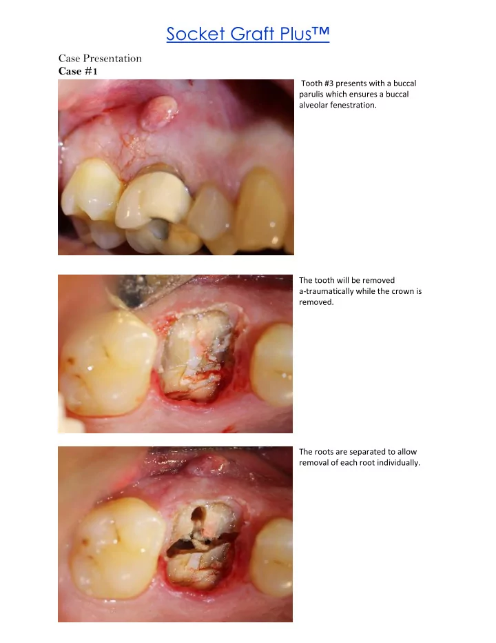
Case Presentation Case #1
Tooth #3 presents with a buccal parulis which ensures a buccal alveolar fenestration. The tooth will be removed a-traumatically while the crown is removed. The roots are separated to allow removal of each root individually.
SLIDE 2
With the tooth and granulation tissue removed, there is an opening into the sinus associated with the palatal root and a buccal fenestration. The graft material is carried to the socket and condensed with a bone graft packer.
SLIDE 3 Case # 2
The following patient is 80 years old. Tooth #12 was extracted and grafted with Socket Graft™ in 2008 and the implant was placed 9 weeks later. This radiograph was taken in in February 2016 showing the excellent mineralization and maintenance of marginal bone typical of Socket Graft™ over time. Caries and a large endodontic lesion required the removal of #13 therefor, was extracted and grafted with Socket Graft Plus™ Post-op with removal of the
- membrane. It is normal for
some of the granules to be left in the area of the gingiva above the alveolar bone. Any graft material in the gingiva above the bone will not become bone. These granules can be removed with an evacuator or left to exfoliate
SLIDE 4
After evacuating the exposed granules, the remaining graft material is encased in early connective tissue. Two weeks post-op the gingiva is closing over the grafted site. Note, the fibrin clot remains until the epithelium covers over the graft site. There are no granules being pushed out of the graft site. The granules in biocompatible bone grafts remain in the socket. However, the granules from non-biocompatible bone grafts continue to push out of the graft site for up to two months.
SLIDE 5 Case #3
Socket Graft Plus™ greatly simplifies the complexities of socket grafting making it ideal for all socket grafting situations. In this case the bone is missing from the mesial buccal wall. The tooth is removed and the socket is filled with Socket Graft Plus™. Note, the granules are above the bone and surrounding gingiva. These granules will be exposed when the membrane is
- removed. Bone will only grow in the
extraction socket and not in the area of the gingiva. Alveolar ridge 11 weeks after extraction and grafting.
SLIDE 6
Implant placement 11 weeks after extraction and grafting. Healing abutment appointment 11 weeks after implant placement.
SLIDE 7
Case #4
Here is another lower molar with a missing buccal wall. The area of bone loss is associated with the missing buccal wall.
SLIDE 8
This is a view of the socket looking at the crest from the lingual aspect. This is a view of the ridge from the buccal. Socket Graft Plus™ simplifies socket grafting. The material is ideal for simple socket grafting but in situations of apical fenestrations or buccal wall dehiscence’s, the protocol for treating the socket is the same. Simply extract the tooth, remove all granulation tissue and pack the socket with the graft material. There is no need for flaps or subgingival membranes or mixing other materials