SLIDE 1 Nature Neuroscience
Circuit level defects in the developing neocortex of fragile X mice.
- J. Tiago Gonçalves, James E. Anstey, Peyman Golshani, and Carlos Portera-Cailliau
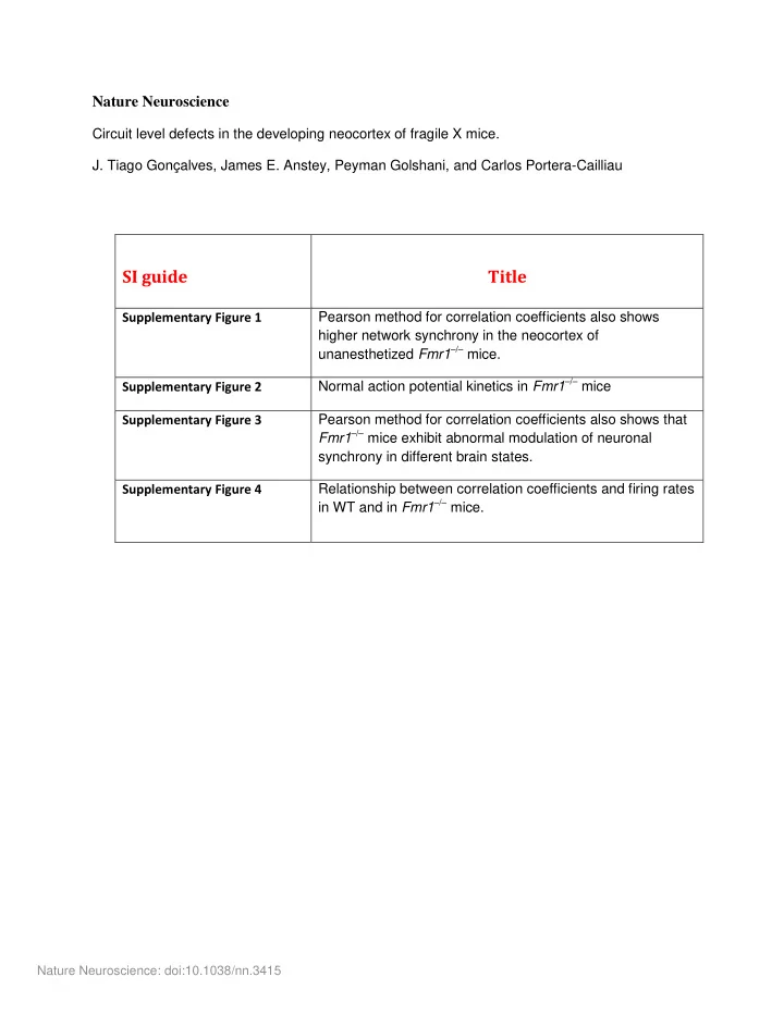
SI guide Title
Supplementary Figure 1 Pearson method for correlation coefficients also shows higher network synchrony in the neocortex of unanesthetized Fmr1–/– mice. Supplementary Figure 2 Normal action potential kinetics in Fmr1–/– mice Supplementary Figure 3 Pearson method for correlation coefficients also shows that Fmr1–/– mice exhibit abnormal modulation of neuronal synchrony in different brain states. Supplementary Figure 4 Relationship between correlation coefficients and firing rates in WT and in Fmr1–/– mice.
Nature Neuroscience: doi:10.1038/nn.3415
SLIDE 2 Mean of Pearson correlations
0.0 0.1 0.2 0.3 0.4 0.5
Correlation coefficient
P9-11 P30-40 P14-16
WT
*
# # # # Fmr1
–/–
Supplementary Figure 1: Pearson method for correlation coefficients also shows higher network synchrony in the neocortex
- f unanesthetized Fmr1–/– mice.
Mean Pearson correlation coefficients for all cell pairs located within 100 µm of each other for WT (black) and Fmr1–/– (red) mice at different postnatal ages. Both age and genotype significantly affect correlation coefficients (#, * Bonferroni corrected p < 0.05, two-way unequal variance ANOVA). The difference in correlation between WT and Fmr1–/– was largest at P14-16 (*p = 0.042, t-test). Error bars indicate the standard error of the mean (s.e.m.). Nature Neuroscience: doi:10.1038/nn.3415
SLIDE 3 0.0 1.0 2.0 3.0 4.0 FHWM (ms) Max fall rate/ Max rise rate
WT WT
2 ms 20 mV
WT
a b c
Fmr1
–/–
Fmr1
–/–
Fmr1
–/–
Supplementary Figure 2: Normal action potential kinetics in Fmr1–/– mice. (a) Sample action potential traces from representative in whole-cell recordings of L2/3 neurons in unanesthetized WT (gray) and Fmr1–/– mice (red) showing similar kinetics (23 and 63 action potentials, respectively). (b, c) Quantification of the (a) Maximum Fall Rate / Maximum Rise Rate ratio (0.26 ± 0.01, n = 7 cells vs. 0.25 ± 0.01 ms, n= 12 cells, p = 0.80, t-test) and (b) full width half maximum (2.17 ± 0.09 ms, n = 7 cells vs. 2.14 ± 0.18 ms, n= 12 cells, p = 0.93, t-test) for action potential traces in WT mice (black) and Fmr1–/– mice (red). Each symbol square/circle represents the average for a different mouse. Nature Neuroscience: doi:10.1038/nn.3415
SLIDE 4
Pearson's Correlations P14-16
0.00 0.05 0.10 0.15 0.20 0.25
Correlation coefficient L/H power > 200 WT
*
Fmr1
–/–
Supplementary Figure 3: Pearson method for correlation coefficients also shows that Fmr1–/– mice exhibit abnormal modulation of neuronal synchrony in different brain states. Mean estimated Pearson correlation coefficients for EEG recordings with L/H power > 200. (WT: 0.114 ± 0.015, n = 6 recordings vs. Fmr1–/–: 0.171 ± 0.012, n = 5 recordings, *p = 0.021, t-test). Nature Neuroscience: doi:10.1038/nn.3415
SLIDE 5
L/H > 200 L/H < 200 p= 0.46 r2 =0.005
** p < 0.01
r2 =0.6462
L/H = 193 L/H = 204
Firing rate vs. correlation coefficient P14-16
Extrapolated firing rate (Hz) Correlation coefficient 0.25 0.15 0.20 0.10 0.05 0.00 0.15 0.25 0.20 0.30 0.35
a b
Firing rate vs. brain state P14-16
L/H <200
(More awake)
L/H >200
(More asleep)
0.0 0.1 0.2 0.3 Extrapolated firing rate (Hz) WT Fmr1
*
–/–
Supplementary Figure 4: Relationship between correlation coefficients and firing rates in WT and in Fmr1–/– mice. (a) Plot of mean Pearson correlation coefficients and firing rates (extrapolated from Ca2+ traces) for WT and Fmr1–/– mice at P14-16. Whereas in WT mice the two were not correlated, in Fmr1–/– mice correlation coefficients increased with higher extrapolated firing rates, which corresponded to mice with L/H Power > 200. (b) Firing rate vs. brain state. Note that, just like with the electrophysiology data (Fig. 3e), firing rates extrapolated from calcium imaging data were higher in Fmr1–/– mice compared to WT mice when mice were more asleep (EEG showing L/H power > 200, WT: 0.222 ± 0.010, n= 6 recordings vs. Fmr1–/–: 0.273 ± 0.017, n = 5 recordings, Bonferroni corrected p = 0.527, two-way ANOVA) but not when mice were more awake (LH power < 200; WT: 0.236 ± 0.013, n= 7 recordings vs. Fmr1–/–: 0.212 ± 0.019, n = 5 recordings, Bonferroni corrected *p = 0.047, two-way ANOVA). There were also non-significant trends towards higher firing in WT mice with lower L/H power and higher firing in Fmr1–/– mice with higher L/H power, just like with the electrophysiology. Nature Neuroscience: doi:10.1038/nn.3415