
SLIDE 1
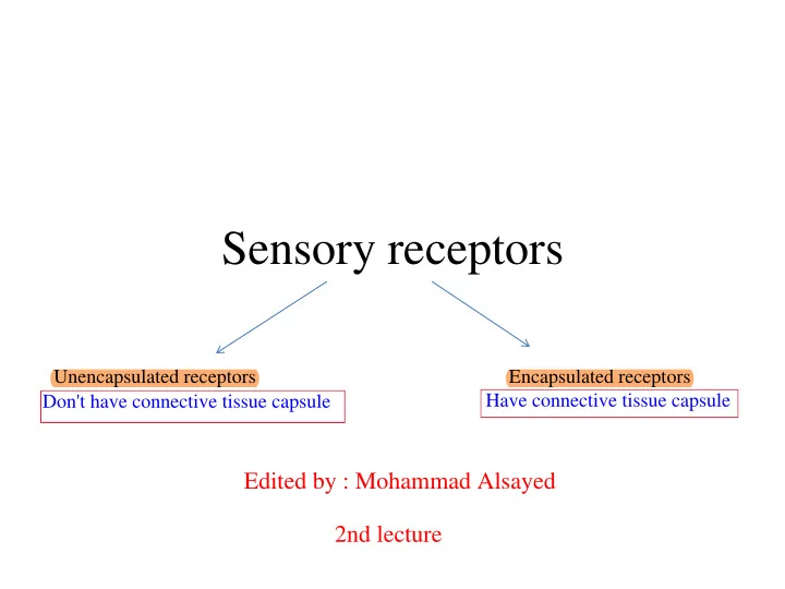
Sensory receptors
Unencapsulated receptors Encapsulated receptors
Have connective tissue capsule
Don't have connective tissue capsule
Edited by : Mohammad Alsayed 2nd lecture
Sensory receptors Unencapsulated receptors Encapsulated receptors - - PowerPoint PPT Presentation
Sensory receptors Unencapsulated receptors Encapsulated receptors Have connective tissue capsule Don't have connective tissue capsule Edited by : Mohammad Alsayed 2nd lecture 1- Merkel disc Unencapsulated nerve receptors for light touch
Unencapsulated receptors Encapsulated receptors
Have connective tissue capsule
Don't have connective tissue capsule
Edited by : Mohammad Alsayed 2nd lecture
1- Merkel disc
merkel cell 2- Free nerve endings
3- Root hair plexuses
reticular dermis
Root hair plexuses
Free nerve endings Merkel disc
sensation produced by pressure receptors in the skin
when they activated
Meissner corpuscles:
soles
Pacinian corpuscles
hypodermis
touch) and vibrations
because it is located superficially You need deep pressure to activate these receptors
Ruffini corpuscles:
Located in the reticular layer of dermis
Hair Follicles and hair Sweat glands Sebaceous glands Nails
The accessory structures that are associated with the skin and formed the ( Integumentary system ) 1) 2) 3) 4)
Hairs are elongated keratinized structures that form within epidermal invaginations (hair follicles) Hair shaft: The part of a hair extending beyond the skin surface (visible part) Hair root: The part of a hair below the skin surface (embedded part) Types of hair: 1- Lanugo: fetal hair 2- Down hair: light colored hair
3- Terminal (adult) hair: thicker, darker hair that begins to grow at puberty
above the skin surface
Hair follicle is a tube of stratified squamous epithelium, invaginated into the dermis INNER ROOT SHEATH Disintegrates at the level of the sebaceous gland OUTER ROOT SHEATH
epidermis
hair formation
basement membrane
surrounded by a connective tissue sheath.
Only stratum basale and stratum spinosum make invagination and form :
Inner root sheath
Hair matrix
that generate the hair and the internal root sheath
papilla
matrix produce hair color.
Inner root sheath reach this level and stops at the
Invagination of the dermis deep inside the hair bulb and it is called (hair papilla) Why the hair is very tough ? because the keratin is hard type and highly compacted , while the keratin in the stratum corneum is softer than keratin in the hair shaft
Sebaceous glands
called sebum, to lubricate and waterproof the skin and hair
secretion
The whole cell is die and secrete the sebum ﺔﻤﻋﺎﻧ ةﺮﺸﺑ كﺪﻨﻋ نﻮﻜﯾ نﺎﺸﻋ ﻢﮭﺗﺎﯿﺤﺑ اﻮﺤﻀﺑ ﺎﯾﻼﺨﻟا لوﺬھ ﮫﻧﺄﻛ ﻲﻨﻌﯾ * In the last lecture we said that the thick skin in the soles and palms does not contain hair , so we do not have sebaceous gland Why ?? Simply , if we have sebaceous gland in the palms or soles , it would be very difficult for you to hold things and you can't walk
* The secretion of this gland is stimulated by : Estrogen + Testosterone . This takes place around puberty time , we start producing these hormones and the skin starts to be oily . * Sometimes the opening (duct) of this gland is "blocked" due to excessive secretion , so the sebum collected below the level
See the next slide :) Simple branched acinar gland
Acne Comedo (blackheads) A comedo is a clogged hair follicle (pore) in the skin. Keratin combines with oil to block the follicle
Sometimes this sebum got infected by "Propionibacterium acne" Remember microbiology :) So acne is common during puberty time . داﺪﺴﻧا
Arrector pili muscles are small muscles extend from hair follicles to the dermal papilla
hairs to stand on end (goose bumps)
system (sympathetic ) Depilatory
Pili = Hair because it is smooth muscle The process that the hair is removed is called (Depilatory) .
Pulls hairs upright when cold or frightened
The attachments of these muscles to dermal papillae cause dimples seen in goosebumps
The role of this muscle is more prominent in animals , because it reduces heat loss through the surface of their bodies
Medulla: large vacuolated and moderately keratinized cells Cortex: heavily keratinized and densely packed cells Cuticle: thin layer heavily keratinized squamous cells covering the cortex
عﺎﺨﻨﻟا ةﺮﺸﻘﻟا and contains pigment melanin very hard
Hairs grow discontinuously, with periods of growth followed by periods of rest and this growth does not occur synchronously in all regions of the body or even in the same area
1) 2) 3) 1) Anagen phase (Active growth phase) : Some people have difficulty growing in their hair , because they have short active growth phase and the opposite is true . Also the type of the hair plays role , like the hair of the arms , legs and eyebrows , all of them have very short active growth phase .
During this phase the dermal papilla starts to disintegrate , starts separation between the hair bulb and dermal papilla , this leads to shrinkage of the hair follicle because there is no blood supply The dermal papilla is completely detached from the hair bulb and hair follicle . Shedding of hair can takes place because no longer attached to dermal papilla There is new formation of dermal papilla within the hair follicle and formation of new hair
85% of hair follicles are in 15 % of hair follicles are in
Medulla Cortex Cuticle Outer root sheath Inner root sheath Epidermis of skin Dermal papilla
ءﺰﺠﻟا ﮫﻧﻷ ﻲﻤﻠﻋ ﺶﻣو ﺔطاﺮﻓ ﻲﻜﺣ دﺎھ ,, ﺔﻤﯾﺮﺟ ﻞﺤﻟ DNAلا اﻮﻠﻠﺤﯾ نﺎﺸﻋ " قرزﻷﺎﺑ دﺪﺤﻤﻟا ءﺰﺠﻟا " ﺮﻌﺸﻟا ﺔﻠﺼﺧ اوﺬﺧﻮﯾ مﻼﻓﻷﺎﺑ ﺎﻤﻟ : ﺶﻣﺎﮭﻟﺎﻋ ﺔﻣﻮﻠﻌﻣ ): .. DNA ﻲﻓ ﺎﻣ ﻲﻟﺎﺘﻟﺎﺑو ةاﻮﻧ ﺎﮭﯿﻓ ﺎﻣ ﺎﯾﻼﺧ ﻦﻋ ةرﺎﺒﻋ ﺮﻌﺸﻟا ﻦﻣ زرﺎﺒﻟا
Dermal papilla
invaginate in the epidermis)
Matrix cells
Melanocytes
59
nutrient source for bacteria (odor !!)
stimulated during emotional distress
abundant on palms & soles: ~ 500/cm2)
H2O; rest NaCl + some waste products
Scent glands
then to skin surface True sweat and forehead The bacteria starts to grow and produce smell with the sweat Not true sweat
Apocrine sweat glands Eccrine (merocrine) sweat glands
62
Small in size , narrower lumen Large in size , wider lumen
Hard plates of keratin on the dorsal surface of each distal phalanx Lack of pigment makes them colorless
Nail parts
cut
area
nail fold (cuticle)
free edge where dirt accumulates
the pink part
, simply because they don't have Melanocytes
and spinosum ﻞﻜﺸﻟا ﺔﯿﻟﻼھ
Nail matrix
Layer of cells at the base of the nail , it consist of rapidly dividing skin cells that soon will fill up with keratin and when the matrix cells undergo mitosis , they push the nail plates over nail beds and that's how the nails grow
Epidermal ridge Dermal papilla
Sebaceous gland Hair follicle Arrector pili Pacinian corpuscle Sweat gland Hair shaft/root Dermal papilla Hair matrix
Junction between dermis an hypodermis
Meissner corpuscle
Pacinian corpuscles
This is the sensory neuron and the white material is the capsulated connective tissue important for deep pressure detection
Sebaceous gland Hair follicle Arrector pili
What type of skin this section ?? Thick skin , because there is stratum lucidum from the palms and the soles . Don't forget the criterias of classification that's we taken in first lecture . A : stratum basale B : stratum spinosum C : stratum granulosum D : stratum lucidum E : stratum corneum
THICK OR THIN SKIN ????
Once you see lucidum layer , that's mean this section is taken from thick skin (ex: palms and soles)