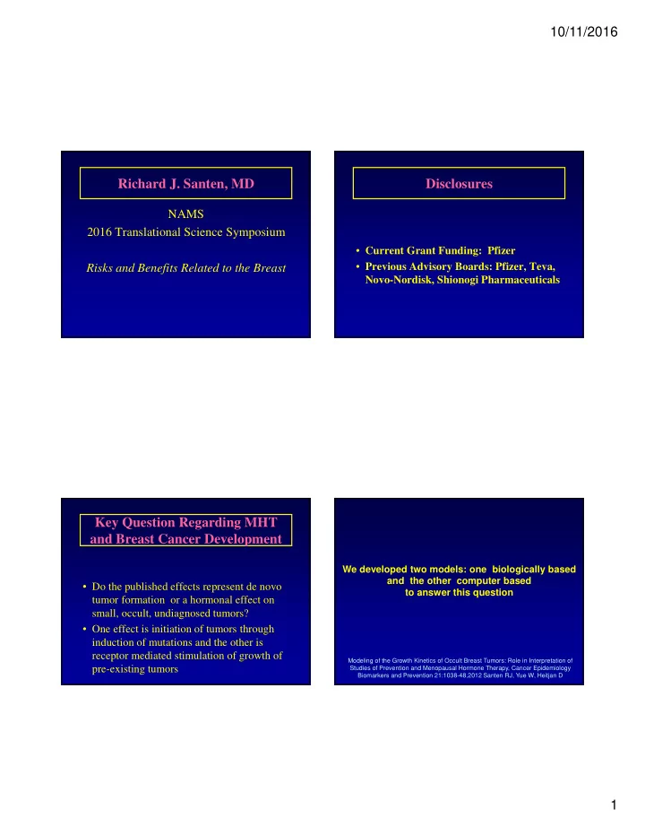

10/11/2016 Richard J. Santen, MD Disclosures NAMS 2016 Translational Science Symposium • Current Grant Funding: Pfizer • Previous Advisory Boards: Pfizer, Teva, Risks and Benefits Related to the Breast Novo-Nordisk, Shionogi Pharmaceuticals Key Question Regarding MHT and Breast Cancer Development We developed two models: one biologically based and the other computer based • Do the published effects represent de novo to answer this question tumor formation or a hormonal effect on small, occult, undiagnosed tumors? • One effect is initiation of tumors through induction of mutations and the other is receptor mediated stimulation of growth of Modeling of the Growth Kinetics of Occult Breast Tumors: Role in Interpretation of pre-existing tumors Studies of Prevention and Menopausal Hormone Therapy, Cancer Epidemiology Biomarkers and Prevention 21:1038-48,2012 Santen RJ, Yue W, Heitjan D 1
10/11/2016 Average of 11 driver Life History of a Breast Tumor mutations in breast cancers IBC IBC Start of mutation Start of mutation cascade through IBC cascade through IBC Initiation events Initiation events DCIS DCIS ADH ADH HELU HELU To be diagnosed, the tumor must exceed the detection threshold Detection threshold What determines the detection threshold? IBC DCIS ADH HELU 2
10/11/2016 Influence of Age Change in mammographic density with age on Detection Threshold • <40 1.63 cm • 40-49 1.44 cm • 50-59 1.25 cm • 60-69 1.07 cm • >70 0.88 cm Average for the WHI age 50-69 1.16 cm 70 80 30 40 50 60 Change in mammographic density with age Change in mammographic density with age 30 40 50 60 70 80 30 40 50 60 70 80 3
10/11/2016 How long does it take for a de novo tumor to reach the It takes 30 doublings for a tumor to go from one cancer detection threshold? Limit of clinical cell to a tumor of a billion cells, the number needed to detection reach a size of 1 cm in diameter IBC DCIS ADH HELU Depends on the doubling time Median approximates 200 days 4
10/11/2016 50 100 150 200 250 How many de novo tumors would have reached the diagnostic Average threshold within the 5.6 year 16 years duration of the WHI E+P study ? 50-69 year old 50 100 150 200 250 Only tumors with a doubling time of 50 days or less 50-69 year old 5
10/11/2016 Percentage of tumors in the WHI arising de novo • 15% of tumors have doubling time of 50 days or less • Only tumors arising de novo during years one and two of the study would have 4.1 years to reach threshold of detection • Years 1-2 detectable; years 3-5 not detectable; 2/5 x 15% = 6% • Therefore only 6% of tumors arose de novo 15% have doubling times Of 50 days or less • The other 94% arose from tumors in the occult, small undiagnosed pool Occult breast cancers diagnosed at autopsy Ages 40-80 What was prevalence of undiagnosed tumors at start of WHI Study? IBC DCIS ADH In Situ 6% Invasive 1% Total 7% HELU 6
10/11/2016 Prevalence of undiagnosed tumors Iterative Modeling at start of WHI Study • Parameters – Doubling time of occult tumors – Percentage of tumors in the reservoir – Detection threshold • Assumptions IBC DCIS – Log linear growth kinetics (confirmed in ADH xenografts; Hormones and Cancer 2013) HELU – Gaussian distribution of sizes of occult tumors in the reservoir De Novo Tumor IBC DCIS ADH HELU 7
10/11/2016 Compare observed with expected 200 day doubling time Observed incidence is taken from the SEER 1998 to 2007 population data Model parameters based on best fit with observed data • 200 day average doubling time • 1.16 cm detection threshold • 7 % prevalence in women ages 50-80 8
10/11/2016 Population incidence Contralateral cancer Computer based model incidence • Calculate yearly incidence of de novo tumors based on age related population incidence data • Vary doubling times based on Gaussian distributions • Correct for deaths from competing causes in the population • Calibrate based on SEER population incidence data Model Developed to interpret risk of breast cancer from MHT 9
10/11/2016 WHI E+P Study Iterations • Used biologically based model – 200 day doubling time Effective doubling times of: – 7% prevalence 190 days – 1.16 cm detection threshold 170 days • 80% of tumors diagnosed in E+P trial were 150 days ER + 130 days 110 days Life history of a breast tumor 150 day doubling time E+P HR 1.26 200 day doubling time IBC DCIS ADH HELU 10
10/11/2016 Hormone naïve Hormone therapy 7 Percent in each doubling 6 5 category 4 3 2 1 0 1 5 10 15 20 25 30 Doublings 41 How does the predicted risk of breast cancer Influence excess risk attributable to E + P ? 43 11
10/11/2016 Excess Risk per Relative Risk Absolute Risk 1000 women Per 5 years per 5 years 1.25 1.0% 3 CEE alone arm 1.25 2.5% 6 of the WHI 1.25 5.0% 13 1.25 7.5% 19 1.25 10% 25 HR 0.77 (CI 0.62-0.95) placebo Hypothesis 23% reduction In breast cancer incidence CEE • Conjugated equine estrogens caused Ages 50-79 apoptosis of occult tumors • Long term deprivation of estrogen causes breast cancer cells to undergo apoptosis in response to estrogen • The average age of women in the WHI was 63, 12 years after the average age of menopause 12
10/11/2016 In Vitro Model of Long Term Wild Type Cells Estrogen Deprivation 1.5 Apoptosis (fold of induction) 1 MCF-7 LTED 0.5 >6 months 0 control -14 -12 -10 -8 E2 concentration (M) Estrogen deprived media Long term anti-estrogen treated xenografts LTED Cells 10 Control Apoptosis (fold of induction) Average tumor volume (cm 2 ) 0.3 cm E 2 8 0.6 6 Treatment Begins 0.4 4 2 0.2 0 P <.0001 control -14 -12 -10 -8 0.0 E2 concentration (M) 1 2 3 4 5 6 7 8 Weeks Data of VC Jordan 13
10/11/2016 P <.00014 60 * Percent apoptosis 50 40 Model based on apoptosis used to predict effect 30 of estrogen alone on breast cancer risk 20 10 0 control E 2 (5 days) Jordan et al Data in support of need for long term estradiol deprivation to experience breast cancer reduction Women treated with estrogen, washed out, and then randomized to CEE experienced no decrease in breast cancer HR 1.02 (0.70-1.50) 14
10/11/2016 Implications Historical Footnote • Need to treat these occult lesions before they become • High dose estrogen was used to treat clinically detectable metastatic breast cancer • A form of hormone therapy for menopausal women which prevents these occult lesions from growing but • Only effective in women at least 5 years relieves menopausal symptoms would be ideal postmenopausal • Proof of principle • Recent studies indicate that physiologic – Tamoxifen prevented breast cancer in a large British trial doses of estradiol also cause tumor even when estrogen was given at the same time to treat regression in 30% of postmenopausal menopausal symptoms – Need to exploit this concept but develop more effective women with metastatic breast cancer methods Thank you for your attention Summary • Menopausal hormone therapy does not cause breast cancer but instead, exerts effects on undiagnosed, occult tumors – E+P enhances tumor growth ( doubling time 200 days to 150 days on average) – E alone given years after menopause causes apoptosis and results in decreased tumor incidence • Breast cancer prevention with tamoxifen or raloxifene represents early treatment not true prevention University of Virginia Richard J. Santen MD 15
Recommend
More recommend