Rheumatology vs Ortho: How Do You Tell? Andrew J. Gross, MD - - PowerPoint PPT Presentation

rheumatology vs ortho how do you tell
SMART_READER_LITE
LIVE PREVIEW

Rheumatology vs Ortho: How Do You Tell? Andrew J. Gross, MD - - PowerPoint PPT Presentation

Disclosures None Rheumatology vs Ortho: How Do You Tell? Andrew J. Gross, MD Rheumatology Clinic Chief Associate Clinical Professor University of California, San Francisco Objectives Clinical Case #1 Recognize the key features of


slide-1
SLIDE 1

1

Rheumatology vs Ortho: How Do You Tell?

Andrew J. Gross, MD Rheumatology Clinic Chief Associate Clinical Professor University of California, San Francisco

Disclosures

  • None

Objectives

  • Recognize the key features of polymyalgia

rheumatica

  • Recognize inflammatory back pain
  • Know the differential diagnosis of subacute

monoarticular arthritis

  • Know the hallmarks of fibromyalgia.
  • Distinguish rheumatoid arthritis from
  • steoarthritis by hand joint involvement
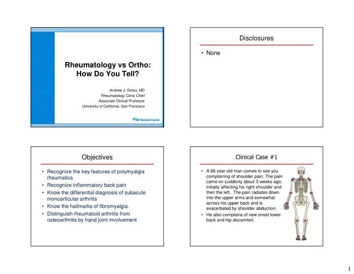
Clinical Case #1

  • A 66 year old man comes to see you

complaining of shoulder pain. The pain came on suddenly about 3 weeks ago, initially affecting his right shoulder and then the left. The pain radiates down into the upper arms and somewhat across his upper back and is exacerbated by shoulder abduction.

  • He also complains of new onset lower

back and hip discomfort.

slide-2
SLIDE 2

2 Clinical Case #1 - Question

You diagnose him with Polymyalgia Rheumatica (PMR). All of the following symptoms tipped you off to the diagnosis of PMR EXCEPT:

  • a. Morning stiffness lasting >45 minutes
  • b. Hand numbness
  • c. Pain & stiffness affects the lower back and pelvic

girdle

  • d. Pain & stiffness improves with activity
  • e. ESR >40 mm/hr

Dasgupta B, et al, Ann Rheum Dis 2012, EULAR/ACR Classification Criteria

All of the following symptoms tipped you off to the diagnosis of PMR EXCEPT:

  • a. AM stiffness >45 min
  • b. Hand numbness
  • c. lower back stiffness
  • d. Better w/ activity
  • e. ESR >40 mm/hr

A M s t i f f n e s s > 4 5 m i n H a n d n u m b n e s s l

  • w

e r b a c k s t i f f n e s s B e t t e r w / a c t i v i t y E S R > 4 m m / h r

3% 67% 9% 19% 2%

Clinical Case #1 - Question

You diagnose him with Polymyalgia Rheumatica (PMR). All of the following symptoms tipped you off to the diagnosis of PMR EXCEPT:

  • a. Morning stiffness lasting >45 minutes
  • b. Hand numbness
  • c. Pain & stiffness affects the lower back and pelvic

girdle

  • d. Pain & stiffness improves with activity
  • e. ESR >40 mm/hr

Dasgupta B, et al, Ann Rheum Dis 2012, EULAR/ACR Classification Criteria

Some Tips about PMR

  • Typical distribution of

PMR symptoms…

  • Subdeltoid bursitis &

biceps tenosynovitis are common in one or both shoulders

  • Patients may develop

adhesive capsulitis

Salvarani, C, et al, Nat Rev Rheumatol, 2012, PMID 22825731

slide-3
SLIDE 3

3 Some more Tips about PMR

  • PMR is uncommon in patients < 60 years old

97 cases of PMR identified during a 10 year study from Olmstead County, Minnesota 0-49 years 1 in a million 50-59 years 1 in 5,000 60-69 years 1 in 2,000 70-79 years 1 in 900

Chuang TY , et al, Ann Intern Med 1983, PMID 6982645

Some more Tips about PMR

  • PMR is uncommon in patients < 60 years old
  • ESR is helpful - but it is <40 mm/hr in 10-20% of patients

– CRP can be helpful when ESR is <40

  • 15% will have Giant Cell Arteritis (new onset head pain)

– New onset head pain – Scalp tenderness – Jaw claudication when chewing – Sudden vision loss or diplopia

Dasgupta B, et al, Ann Rheum Dis 2012, EULAR/ACR Classification Criteria

Things patients with PMR often tell me

  • “I feel like I am 100 years old!”
  • “I need to crawl out of bed in

the morning”

  • “I feel okay as long as I keep

moving, but I stiffen up as soon as I sit down – like the tin man”

  • “That prednisone is a miracle”

When To Refer PMR to a rheumatologist

  • Rheumatologists are generally pleased to see cases
  • f PMR
  • Partial response to treatment with prednisone –

should have fully response to 15-20 mg/d.

  • Difficulty tapering prednisone
  • Symptoms of Giant Cell Arteritis
slide-4
SLIDE 4

4 Clinical Case #2

  • A 26 year old man comes to see you

complaining of shoulder pain. The pain came on about 3 weeks ago, initially affecting his right shoulder and then the left. The pain does not radiate. Range of motion of motion of both shoulders is limited.

  • He also notices pain and stiffness in

his neck and lower back. This is worse recently, but has been present

  • n an off for the past couple of years.
  • He complains of a hour of morning

stiffness in his shoulders and low back.

Clinical Case #2

  • The shoulder exam is notable for

limitation in shoulder ROM (abduction, internal & external rotation) without weakness in the rotator cuff muscles. There is some tenderness over the glenohumeral joint. No effusion.

  • Cervical spine flexion & rotation as

well as lumbar spine flexion are somewhat limited. Straight leg raise is unremarkable.

  • Hip rotation is also somewhat limited.
  • The remainder of the joint exam is

unremarkable.

Clinical Case #2

Which of the following conditions is the most likely cause of this man’s shoulder, neck and lower back pain:

  • a. Ankylosing Spondylitis
  • b. Polymyalgia Rheumatica
  • c. Rheumatoid Arthritis
  • d. Systemic Lupus Erythematosus
  • e. Calcium Pyrophosphate Dihydrate Disease

(CPPD) Which of the following conditions is the most likely cause of this man’s shoulder, neck and lower back pain:

  • a. AS
  • b. PMR
  • c. RA
  • d. SLE
  • e. CPPD

A S P M R R A S L E C P P D

82% 4% 1% 1% 10%

slide-5
SLIDE 5

5

Clinical Case #2

Which of the following conditions are a likely cause of this 26 y.o. man’s shoulder, neck and lower back pain:

  • a. Ankylosing Spondylitis
  • b. Polymyalgia Rheumatica (age >50)
  • c. Rheumatoid Arthritis
  • d. Systemic Lupus Erythematosus
  • e. Calcium Pyrophosphate Dihydrate Disease

(CPPD) (usually older people, typically spares lumbar spine)

Typical distribution

  • f involved joints in

rheumatoid arthritis (and lupus)

www.studyblue.com

https://dundeemedstudentnotes.wordpress.com/2014/06/16/polyarthritis/

Rheumatoid Arthritis Psoriatic Arthritis Ankylosing Spondylitis Osteoarthritis

Ankylosing Spondylitis

slide-6
SLIDE 6

6 Ankylosing Spondylitis - sacroiliitis AS – “bamboo spine”

Back pain Sacroiliitis on MRI Back pain Radiographic sacroiliitis Back pain

Syndesmophytes

DIAGNOSIS

Ankylosing Spondylitis

Rudwaliet M, et al. Arthritis Rheum. 2005;52(4):1000-1008.

Non-radiographic stage Radiographic stage

Time (years)

Clinical Case #2

All of the following symptoms are associated with inflammation of the spine (spondylitis) EXCEPT:

  • a. Pain & stiffness improve with exercise.
  • b. Onset of back pain was insidious
  • c. Back pain & stiffness gets worse at night
  • d. Burning pain in the thighs with standing
  • e. Symptoms began before age 40
slide-7
SLIDE 7

7

All of the following symptoms are associated with inflammation of the spine (spondylitis) EXCEPT:

  • a. Improves w/ exercise
  • b. Insidious onset
  • c. Pain worse at night
  • d. Burning pain in thighs
  • e. Symptoms <40 y.o.

I m p r

  • v

e s w / e x e r c i s e I n s i d i

  • u

s

  • n

s e t P a i n w

  • r

s e a t n i g h t B u r n i n g p a i n i n t h i g h s S y m p t

  • m

s < 4 y .

  • .

14% 7% 4% 62% 14%

Clinical Case #2

All of the following symptoms are associated with inflammation of the spine (spondylitis) EXCEPT:

  • a. Pain & stiffness improve with exercise.
  • b. Onset of back pain was insidious
  • c. Back pain & stiffness gets worse at night
  • d. Burning pain in the thighs with standing
  • e. Symptoms began before age 40

Inflammatory Back Pain: Hallmark Features

Feature Odds Ratios Insidious onset 12.7 Pain at night (with improvement upon getting up) 20.4 Age at onset <40 years 9.9 Improvement with exercise 23.1 No improvement with rest 7.7 Sensitivity 79.6% & Specificity 72.4% Positive LR = 79.6/(100-72.4) = 2.9 ~ Probability = 14%

Sieper J, et al, Ann Rheum Dis 2009, PMID 19147614 Rudwaleit M, et al. Ann Rheum Dis. 2009; 68(6):777-83. Ozgocmen S, et al. J Rheumatol. 2010;37(9):1978.

LR=likelihood ratio

NHANES 2009-2010

  • 19.2% of US Adults age 20-69 years old reported

chronic axial pain

  • In patients with chronic axial pain, 28% to 35.5%

had Inflammatory Back Pain

  • US prevalence of IBP: 5% to 6%

Reveille JD, et al. Arthritis Care & Res. 2012;64(6):905-910.Weisman MH, et al. Ann Rheum Dis. 2013;72(3):369-373.

  • Self-reported prevalence of ankylosing spondylitis = 0.55
slide-8
SLIDE 8

8 When to refer a patient with back pain to a rheumatologist

  • Inflammatory Back Pain
  • HLA-B27+ (present in 85-95% of patients with AS)
  • Elevated CRP
  • Sacroiliitis on imaging (x-rays or MR)
  • Family history of Ankylosing Spondylitis

Poddubnyy D, van Tubergen A, Landewé R, et al. Ann Rheum Dis 2015;74:1483–1487

AS: Treatment

NSAID NSAIDs sulfasalazine TNF inhibitors

Axial disease only

Physical Therapy

Braun J, et al.,, Ann Rheum Dis 2011; 70: 896-904; van der Heijde D, et al, Ann Rheum Dis 2011; 70:905-08

Clinical Case #3

  • 45 year old man comes to see you with left knee

swelling for the past 7 days. He has no other complaints.

  • ROS is unremarkable. No fevers or rashes
  • Physical Exam: unremarkable except for swelling

and warmth of the left knee with limited ROM.

Clinical Case #3

To identify the cause of the knee swelling, what is the best next test to obtain:

  • A. Aspirate Knee Fluid for cell count and crystal search
  • B. MRI of knee
  • C. X-ray of knee
  • D. CBC with Differential
  • E. Rheumatoid factor & CCP antibody
slide-9
SLIDE 9

9 To identify the cause of the knee swelling, what is the best next test to obtain

  • A. Aspirate Knee Fluid
  • B. MRI of knee
  • C. X-ray of knee
  • D. CBC w/ diff
  • E. RF & CCP

A s p i r a t e K n e e F l u i d M R I

  • f

k n e e X

  • r

a y

  • f

k n e e C B C w / d i f f R F & C C P

59% 6% 9% 3% 24%

Differential Diagnosis of Sub-Acute Monoarticular Arthritis

Non-Inflammatory

  • Cartilage or ACL tear
  • Flare of osteoarthritis
  • Mimics of joint swelling

– Prepatellar bursitis – Body habitus (adipose tissue) and tendinitis

Inflammatory

  • Infectious

– Lyme Disease – Gonococcus

  • Crystal

– CPPD – Gout

  • Autoimmune

– Spondyloarthritis – Palindromic rheumatism – Other systemic disease

Aspirate the Knee! Synovial Fluid Analysis Cell Count & Crystal Search

Quest Diagnostics

  • Test Code 4707

LabCorp

  • Test Code 005231
  • Green top tube preferred

(lavender top tub will work)

  • 1-10 cc
  • CPT: 89051; 89060
  • Refrigerated (do not freeze)
  • Okay for up to 2 days

Zuber TJ, Am Fam Phys 2002 www.aafp.org/afp/2002/1015/p1497.html

Synovial Fluid Analysis Cell Count & Crystal Search

Zuber TJ, Am Fam Phys 2002 www.aafp.org/afp/2002/1015/p1497.html

Type Non- Inflammatory e.g.

  • steoarthritis

Inflammatory e.g. rheumatoid arthritis Infectious e.g. crystal or septic Appear- ance Clear Viscous amber Turbid yellow less viscous Turbid yellow less viscous WBC <2000 cells/mm3 2000 - 50,000 cells/mm3 >50,000 cells/mm3 Cell Type Mononuclear PMNs and/or lymphocytes PMNs

slide-10
SLIDE 10

10 Synovial Fluid Analysis Cell Count & Crystal Search

Zuber TJ, Am Fam Phys 2002 www.aafp.org/afp/2002/1015/p1497.html

Tips on subacute septic arthritis

Erythema Chronicum Migrans

Tips on subacute septic arthritis

Lyme Disease

  • Unlikely unless traveled to

Lyme endemic region

  • Initial phase with erythema

migrans rash & sometimes fever and diffuse arthralgia

  • If untreated, later can

develop monoarticular arthritis, usually of the knee

  • Lyme ELISA & WB will be

strongly positive

  • No role for testing joint fluid

www.findarthritistreatment.com/eight-causes-of-migrating-arthritis/

Tips on subacute septic arthritis

Lyme Disease

  • Unlikely unless traveled to

Lyme endemic region

  • Initial phase with erythema

migrans rash & sometimes fever and diffuse arthralgia

  • If untreated, later can

develop monoarticular arthritis, usually of the knee

  • Lyme ELISA & WB will be

strongly positive

  • No role for testing joint fluid

Gonococcus

  • Sexually transmitted disease
  • Classically initially presents

with tenosynovitis of the wrist eventually settling in to become a septic joint.

  • Can involve multiple joints
  • Often with scattered pustular

skin rash (easy to miss)

  • DNA testing from urine and

throat swab.

  • No role for culture from

blood or joint fluid.

slide-11
SLIDE 11

11

Forms of Spondyloarthritis

Tips on spondyloarthritis

Reactive arthritis

  • Sterile oligoarticular arthritis,

usually of lower extremities

  • Develops 10-14 days

following an infectious process, usually dysentery

  • r chlamydia urethritis
  • Sometimes associated with

– Conjunctivitis or uveitis – Urethritis (independent of Chlamydia)

  • More than 50% of cases will

resolve in <6 months.

Psoriatic Arthritis

  • Occurs in 15% of patients

with psoriasis

  • More common in people with

psoriasis affecting the scalp

  • r diffuse severe disease

Psoriasis Clinical Case #5

A B D

slide-12
SLIDE 12

12

A B C D

T2 MRI

Case 5: A 50 year old healthy active woman with severe exacerbation of chronic right shoulder pain. Which image is most likely associated with her disorder?

Case 5: A 50 year old healthy active woman with exacerbation of chronic right shoulder pain. Which image is most likely associated with her disorder?

  • A. A
  • B. B
  • C. C
  • D. d

A B C D

A B C D

18% 22% 30% 30%

A B C D

T2 MRI

Case 5: A 50 year old healthy active woman with exacerbation of chronic right shoulder pain. Which image is most likely associated with her disorder?

A B C D

  • A. Rheumatoid arthritis (late disease)
  • B. Milwaukee Shoulder Syndrome (apatite-

associated destructive arthritis)

  • C. Calcific Tendinitis
  • D. Rotator cuff tear

T2 MRI

roentgenrayreader.blogspot. com

slide-13
SLIDE 13

13 Clinical Case #6

49 year old woman with multiple

  • complaints. Most of her complaints

focus on pain at various locations. These problems have been present for 6 months. Pain is particularly intense

  • ver the neck, shoulders, low back,

hips, knees, hands and feet. Pain is severe in the AM and worsens over the course of the day. Activity seems to make the pain worse. She sleeps

  • poorly. She has abdominal pains,
  • ccasional loose stool, and she has

headaches as well. She has pruritic skin rashes that are transient.

Clinical Case #6

Which of the following studies will be most useful in establishing this patient’s diagnosis?

  • A. Antinuclear antibody assay
  • B. Rheumatoid factor assay
  • C. Anti-cyclic citrullinated peptide antibody
  • D. HLA-B27
  • E. No additional studies

Which of the following studies will be most useful in establishing this patient’s diagnosis?

  • A. ANA
  • B. RF
  • C. CCP
  • D. HLA-B27
  • E. None

A N A R F C C P H L A

  • B

2 7 N

  • n

e

17% 6% 56% 6% 14%

Clinical Case #6

Which of the following studies will be most useful in establishing this patient’s diagnosis?

  • A. Antinuclear antibody assay
  • B. Rheumatoid factor assay
  • C. Anti-cyclic citrullinated peptide antibody
  • D. HLA-B27
  • E. No additional studies
slide-14
SLIDE 14

14 Clinical Case #6

49 year old woman with multiple

  • complaints. Most of her complaints

focus on pain at various locations. These problems have been present for 6 months. Pain is particularly intense

  • ver the neck, shoulders, low back,

hips, knees, hands and feet. Pain is severe in the AM and worsens over the course of the day. Activity seems to make the pain worse. She sleeps

  • poorly. She has abdominal pains,
  • ccasional loose stool, and she has

headaches as well. She has pruritic skin rashes that are transient. ANA – Lupus RF & CCP – RA HLA-B27 – AS

Inflammatory vs. Non-inflammatory

  • What is your worst time of day?
  • Duration of AM stiffness?
  • Does the pain/stiffness improve/worsen with

activity?

What are the hallmarks of fibromyalgia? (Choose 3)

a) Widespread pain b) Joint Pain c) Non-restful sleep d) Depression e) Fatigue f) Obesity

W i d e s p r e a d p a i n J

  • i

n t P a i n N

  • n
  • r

e s t f u l s l e e p D e p r e s s i

  • n

F a t i g u e O b e s i t y

What are the hallmarks of fibromyalgia? (choose 3)

a) Widespread pain b) Joint Pain c) Non-restful sleep d) Depression e) Fatigue f) Obesity

slide-15
SLIDE 15

15

Diagnosis of Fibromyalgia

ACR classification criteria – Wolfe F, et al, Arthritis Rheum 1990 ACR diagnostic criteria – Wolfe F, et al, Arthritis Rheum 2010

Widespread Pain

(>3 months)

www.ehow.com/about_5059501_fibromyalgia-diagnosis-symptoms.html

Diagnosis of Fibromyalgia

Widespread Pain

(>3 months)

  • Fatigue
  • Poor Sleep
  • Cognitive

Problems

  • Other Sx

ACR classification criteria – Wolfe F, et al, Arthritis Rheum 1990 ACR diagnostic criteria – Wolfe F, et al, Arthritis Rheum 2010

Tender Point Exam

ACR fibromyalgia classification criteria: pain at 11 of 18 points

Fibromyalgia is present when: WPI score ≥7 WPI 3-6 SS score ≥5 SS score ≥9

Clauw D, 2014, JAMA, PMID 24737367

Wolfe F et al, J Rheumatol 2011, PMID 24737367

  • r
slide-16
SLIDE 16

16

Fibromyalgia

Headache/Migraine TMJ disorder Dermatitis/ pruritis Chronic eye irritation/dryness Atypical chest pain Irritable bowel syndrome Polyuria/frequency (“interstitial cystitis”) Dyspareunia/ vulvodynia Paresthesia Chronic fatigue Muscle Cramps Dypsnea Multiple sensitivities

modified from Aaron LA, et al. Arch Int Med. 2000;160:221-227.

Diagnosis of Fibromyalgia

Widespread Pain

(>3 months)

  • Fatigue
  • Poor Sleep
  • Cognitive

Problems

  • Other Sx
  • ther

disease

ACR classification criteria – Wolfe F, et al, Arthritis Rheum 1990 ACR diagnostic criteria – Wolfe F, et al, Arthritis Rheum 2010

Work-up

  • Laboratory Tests

– ESR, CRP – CBC w/ diff – Comprehensive Metabolic Panel (inc. LFTs, Calcium) – Fasting Glucose – Hepatitis B & C – TSH, free T4 – Vitamin D 25-OH – CPK (if appropriate)

– ANA (rarely)

  • X-rays of affected

areas to investigate joint damage

  • Biopsy tissues that

appear affected (ie. skin rashes)

  • EMG for persistent

neurologic symptoms

Tip offs that suggest your patient may have fibromyalgia:

  • Multiple different pain complains

– Eg. Back pain, knee pain, neck pain

  • Activity has consequences

– Eg. If I do ___, I will be in bed for 2 days

  • No position relieves pain symptoms
  • The person has odd complaints –

“my body feels like it has tinnitus”

  • You walk into the room and the lights

are turned down (hypersensitivity).

  • “Pan-positive review of systems”
  • You feel exhausted after the

interview

slide-17
SLIDE 17

17 Costs Related to a Diagnose FMS and Failure to Diagnose FMS

Annemans et al, Arthritis Rheum 2008; 58:895

Summary

  • Don’t diagnose patients <50 y.o. with PMR
  • Recognize inflammatory back pain
  • Aspirate swollen joints
  • Recognize calcific tendinitis
  • Recognize symptoms of fibromyalgia

Thanks! Treatment of Fibromyalgia

Medicine Mind Body

A multidisciplinary approach

slide-18
SLIDE 18

18 Medications to Treat Fibromyalgia

Modest – Strong Evidence for Efficacy in Fibromyalgia

  • Tricyclic antidepressants

– amitriptyline (Elavil) 25-50mg qHS

  • Dual-reuptake inhibitors (SNRIs)

– milnacipran (Savella) – duloxetine (Cymbalta) – venlafaxine (Effexor)

  • Serotonin reuptake inhibitors (SSRIs)

– Fluoxetine (Prozac) 20-80mg/d

  • gabapentin (Neurontin)
  • pregabalin (Lyrica)
  • cyclobenzaprine (Flexeril)

10-30mg at bedtime

  • tramadol 200-300mg/d

No Evidence for Efficacy

  • NSAIDs (Yunus MB, at al, J Rheum 1989)

(Goldenberg DL, et al, Arthritis Rheum 1986)

  • Corticosteroids (Clark S, et al, J Rheumatol 1985)
  • Benzodiazepenes (Quijada-Carrera J, et al, Pain

1996)

  • Opioids (Sorensen J, et al, Scand J Rheumatol 1995)

Goldenberg DL, Burckhardt C, Crofford L, JAMA 2004 Carville SF, et al, EULAR recommendations, Ann Rheum Dis 2008

Non-Pharmacologic Therapies for Fibromyalgia

Weak Evidence for Efficacy

  • Strength training
  • Acupuncture
  • Hypnotherapy
  • Biofeedback
  • Balneotherapy

No Evidence for Efficacy

  • Trigger point injection

Goldenberg DL, Burckhardt C, Crofford L, JAMA 2004 Carville SF, et al, EULAR recommendations, Ann Rheum Dis 2008

Moderate Evidence for Efficacy

  • Aerobic Exercise (efficacy not

maintained if exercise stops)

  • Cognitive Behavioral Therapy
  • Patient Education
  • Group Therapy

Very Weak Evidence

  • Chiropractic therapy
  • Manual & Massage Therapy
  • Electrotherapy
  • Ultrasound

A Rheumatologist’s Approach to the Musculoskeletal Examination

  • Hands

– Identifying the DIP, PIP, MCP – Pitting of the fingernails – Synovitis vs. Dactylitis – Identifying deformities

  • Heberdon’s & Bouchard’s nodes
  • Finger joint subluxation from OA
  • Subluxation of MCPs from RA

– Trigger finger – stenosing tenosynovitis

  • Wrists

– Wrist vs. CMC – DeQuervain’s

  • Elbow

– Olecranon bursitis – Lateral Epicondylitis – Synovitis of the joint

  • Foot

– Locations for gout including 1st MTP bursa – Achilles enthesitis – Talonavicular arthritis – Hammer toe deformities

slide-19
SLIDE 19

19

Pattern of Joint Involvement

  • A. Osteoarthritis
  • B. Rheumatoid Arthritis
  • C. SLE
  • D. Parvovirus B19 arthritis

All of the following conditions often involve the MCP joints and wrists EXCEPT:

O s t e

  • a

r t h r i t i s R h e u m a t

  • i

d A r t h r i t i s S L E P a r v

  • v

i r u s B 1 9 a r t h r i t i s

0% 0% 0% 0%

10

Pattern of Joint Involvement

All of the following conditions commonly involve MCP joints, wrists and knees EXCEPT:

  • A. Osteoarthritis
  • B. Rheumatoid Arthritis
  • C. SLE
  • D. Parvovirus B19 induced arthritis
  • SLE typically has extra-articular

manifestations (rashes)

  • Viral Arthritis typically resolves in <6 weeks

although Chikungunya can last longer

http://www.mridoc.com/mskatlas/Arthritis/Arthritis_Common_Joints_Involved/

Osteoarthritis

  • Osteoarthritis of the hands is common and

rheumatology consultation is usually not

  • necessary. It can be managed with:

– Acetaminophen 1 gm three times a day – NSAIDs if normal kidney function and no risk factors for gastritis – Topical Diclofenac 1% gel – Hand Therapy – Paraffin baths

See American College of Rheumatology Guidelines - www.rheumatology.org