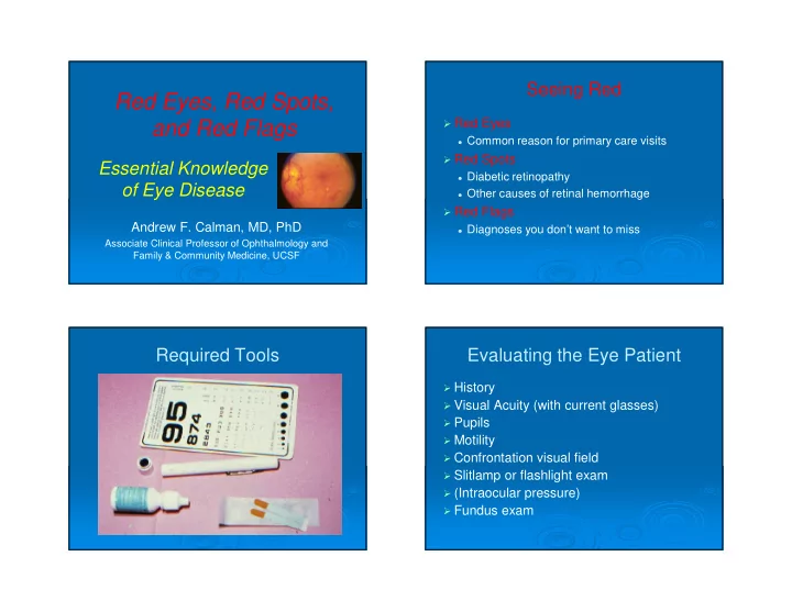
Red Eyes, Red Spots, and Red Flags Red Eyes Common reason for - - PowerPoint PPT Presentation

red eyes red spots and red flags
SMART_READER_LITE
LIVE PREVIEW

Red Eyes, Red Spots, and Red Flags Red Eyes Common reason for - - PowerPoint PPT Presentation

3/25/2013 Seeing Red Red Eyes, Red Spots, and Red Flags Red Eyes Common reason for primary care visits Red Spots Essential Knowledge Diabetic retinopathy of Eye Disease Other causes of retinal hemorrhage Red Flags


slide-1
SLIDE 1

3/25/2013 1

Essential Knowledge

  • f Eye Disease

Andrew F. Calman, MD, PhD

Associate Clinical Professor of Ophthalmology and Family & Community Medicine, UCSF

Red Eyes, Red Spots, and Red Flags

Seeing Red

Red Eyes

Common reason for primary care visits

Red Spots

Diabetic retinopathy Other causes of retinal hemorrhage

Red Flags

Diagnoses you don’t want to miss

Required Tools Evaluating the Eye Patient

History Visual Acuity (with current glasses) Pupils Motility Confrontation visual field Slitlamp or flashlight exam (Intraocular pressure) Fundus exam

slide-2
SLIDE 2

3/25/2013 2

The Red Eye

What is the primary symptom?

Itching and burning Discharge Redness Foreign body sensation Eyelid swelling Pain without discharge

Primary Symptom: Itching and Burning

Blepharitis Allergic Conjunctivitis

Blepharitis

Seborrheic Ulcerative

Acne Rosacea w/Blepharitis

slide-3
SLIDE 3

3/25/2013 3

Blepharitis

Seborrheic – accumulation of desquamated

skin and oils on lids/lashes

Ulcerative – chronic staph colonization Treatment:

Eyelid hygiene: warm compresses, lid scrubs Erythromycin ointment in ulcerative cases Allergy drops if coexisting allergic conjunctivitis Doxy or minocycline if underlying rosacea

Allergic Conjunctivitis Allergic Conjunctivitis

Chronic itching and burning

May be seasonal May be associated with specific allergens

Clinical features

Conjunctiva injected, sometimes edematous Chronic watery or mucoid discharge Numerous papillae on tarsal conjunctiva

(inside the eyelid)

Allergic Conjunctivitis: Tx

Topical medications

Steroids (risk of cataract and glaucoma) Multiple-site agents (olopatidine, OTC

ketotifen)

Antihistamines Mast cell stabilizers (cromolyn sodium) NSAID’s? (diclofenac, ketorolac) Artificial tears

slide-4
SLIDE 4

3/25/2013 4

Primary Symptom: Discharge

Viral conjunctivitis:

Watery discharge (may be thicker in a.m.)

Bacterial conjunctivitis:

Purulent discharge

Allergic conjunctivitis:

Mucoid discharge

Viral Conjunctivitis

Presenting symptoms:

Watery discharge Redness, irritation Acute or subacute onset Often recent URI Usually unilateral Vision only mildly affected May have mild pain and photophobia Etiology: adenovirus, many others

Viral conjunctivitis: Tx

Treatment:

Handwashing to prevent spread Artificial tears Sunglasses when outside Cool compresses Refer if worsening, vision blurred, or if not

resolved in 1-2 weeks

Bacterial Conjunctivitis

Clinical features

Purulent discharge Mild irritation Frequent in pediatric age group Etiology: staph, strep, many others

Treatment

Self-limited: antiobiotic eyedrops are optional

E.g. polymyxin-trimethoprim, gentamicin, sulfacetamide

Refer if severe or persistent, or if signs of eyelid

cellulitis develop

slide-5
SLIDE 5

3/25/2013 5

Primary Symptom: Redness

Subconjunctival hemorrhage Pterygium/pinguecula Episcleritis

Subconjunctival Hemorrhage

Treatment: Reassurance, not referral

Pterygium and Pinguecula Pterygium and Pinguecula

Pinguecula: hyperplasia of sun-damaged

conjunctiva, medial or lateral to limbus

Pterygium: abnormal conjunctiva loses

contact inhibition, partially covers cornea

Treatment:

Eyedrops: antihistamines, vasoconstrictors,

NSAID, avoid steroids

Surgery: excise pterygium, place conjunctival

autograft to prevent regrowth

slide-6
SLIDE 6

3/25/2013 6

Episcleritis Episceritis

Painless dilation of episcleral vessels,

usually in one sector of one eye

Usually benign and self-limited Occasionally associated w/rheum disease Treatment: refer to oph for topical steroids Scleritis: more intense dilation of deep

scleral vessels, severe pain

Primary Symptom: Foreign Body Sensation

Dry Eyes Herpetic Keratitis Foreign Body

Dry Eyes

Clinical presentation

Chronic dryness, irritation or tearing May have associated dry mouth Exam findings subtle

Multiple etiologies

Decreased aqueous secretion with age Unstable tear film due to blepharitis Autoimmune destruction of accessory lacrimal

glands, e.g. in rheumatoid arthritis

slide-7
SLIDE 7

3/25/2013 7

Dry Eyes: Treatment

Treatment:

Tear supplementation Punctal plugs or permanent occlusion Treat associated blepharitis Cyclosporine eyedrops in severe cases

Herpes Keratitis Herpes Keratitis

Clinical presentation

Acute or subacute onset Mild irritation, vision usually normal No discharge (may have mild tearing) Key exam finding: dendritic corneal staining

with fluorescein

Herpes Keratitis

Treatment:

All cases should be referred to ophthalmologist Oral acyclovir (or related compounds) Topical antivirals (trifluorothymidine, ganciclovir)

sometimes used

Topical steroids for deep corneal involvement or

herpetic iritis

Permanent corneal scarring may develop in recurrent

cases

Corneal transplantation sometimes necessary in

severe or recurrent cases

slide-8
SLIDE 8

3/25/2013 8

Herpes Zoster Ophthalmicus Herpes Zoster Ophthalmicus

Vesicular rash in V1 distribution May have keratitis, uveitis, rarely retinitis History of childhood zoster infection Common in elderly and immunosuppressed

patients

Consider HIV test

Treatment: systemic antivirals (aciclovir, etc) Ophthalmology consult to rule out ocular

involvement

Corneal Foreign Body

Foreign Bodies

Speck on cornea or conjunctiva

May be inside eyelid – need to evert lids Remove at slit lamp with foreign body spud Avoid using needles – risk of injury Post-removal antibiotic prophylaxis NSAID drops for pain relief Refer if central or deep

slide-9
SLIDE 9

3/25/2013 9

Primary Symptom: Swelling

Blepharitis (already discussed) Chalazion or hordeolum Preseptal cellulitis Orbital cellulitis Proptosis

Chalazion and Hordeolum Chalazion and Hordeolum

Clinical Presentation

Chalazion: blocked meibomian oil gland with

nontender swelling

Hordeolum: blocked sweat gland with

infection and tender swelling

Chalazion and Hordeolum

Treatment

Hordeolum:

  • Warm compresses, massage
  • Consider systemic and topical antibiotic
  • Monitor for development of preseptal cellulitis

Chalazion:

  • Warm compresses, massage
  • Steroid injection
  • Incision and drainage (from inner aspect of lid)
slide-10
SLIDE 10

3/25/2013 10

Preseptal Cellulitis Orbital Cellulitis Preseptal and Orbital Cellulitis

Preseptal Cellulitis:

Pain and swelling of eyelids Exam: Diffuse lid erythema, edema, tenderness

Orbital Cellulitis: signs of orbital involvement

Proptosis Chemosis (conjunctival edema) Diminished vision, pupil response or motility Fever

Preseptal and Orbital Cellulitis: Tx

Preseptal Cellulitis:

Oral antibiotics, e.g. trimethoprim-sulfa DS II

po bid

Warm compresses Careful monitoring for progression

Orbital cellulitis

CT to rule out orbital abscess IV antibiotics (consider MRSA coverage) Careful monitoring for progression to

cavernous sinus thrombosis or brain abscess

slide-11
SLIDE 11

3/25/2013 11

Contact Dermatitis Contact Dermatitis

Erythema, non-tender edema, itching of eyelids

and face

Most common antigens: eyedrops, cosmetics Treatment:

Identify and remove offending antigen Mild steroid cream/ointment Mild steroid and antihistamine eyedrops if ocular

involvement

Consider systemic antihistamine or steroid if severe

Proptosis Proptosis

Bilateral:

Most common dx: thyroid orbitopathy Check thyroid labs, including Ab’s, and refer

Unilateral

Thyroid still most common etiology Ddx: orbital tumors, inflammatory

pseudotumor, vascular anomalies, myopic degeneration

Check thyroid labs, including Ab’s, and refer

slide-12
SLIDE 12

3/25/2013 12

Red Spots: Diabetic Retinopathy Red Spots: Diabetic Retinopathy

Diabetic retinopathy

Epidemic of preventable blindness Leading cause of blindness in working-age

Americans

Refer all patients for annual dilated exam by

an ophthalmologist

Hypertensive Retinopathy Hypertensive Retinopathy

Hypertensive retinopathy

Fundus findings similar to diabetic retinopathy Not a major cause of vision loss by itself When severe, the tx is to reduce the BP Associated disorders may cause vision loss:

  • Retinal artery occlusion
  • Retinal vein occlusion
  • Ischemic optic neuropathy
  • Occipital stroke
slide-13
SLIDE 13

3/25/2013 13

Diabetic Retinopathy

An epidemic of preventable blindness

At least 90% preventable with proper

screening and treatment

Retinopathy may be present at time of DM dx Retinopathy may be present even with 20/20

vision

By the time patients are symptomatic,

permanent vision loss has occurred

Non-Proliferative Diabetic Retinopathy

Non-Proliferative DR

Microaneurysms (the source of edema) Dot, blot and flame hemorrhages Hard exudates (a sign of edema) Cotton-wool spots (a sign of ischemia) Treatment: usually none at this stage

Optimize glycemic and BP control

Proliferative Diabetic Retinopathy

slide-14
SLIDE 14

3/25/2013 14

Proliferative DR

Hallmark is neovascularization (NV)

Fragile vessels that can leak, bleed and scar May occur on optic disc, retina or iris

Consequences of NV

Vitreous hemorrhage Traction retinal detachment Neovascular glaucoma

Treatment: Panretinal laser, sometimes

vitrectomy, bevacizumab?. Guarded prognosis.

Diabetic Macular Edema Diabetic Macular Edema

Most common cause of vision loss in

diabetics

Detected by stereoscopic biomicroscopy

  • r optical coherence tomography

Leakage sites identified by fluorescein

angiography

Evidence-based criteria for treatment of

“clinically significant” DME

Treatment of DME

Focal laser treatment

Best-studied treatment Validated in Early Treatment of Diabetic

Retinopathy Study

Newer treatments

Injected VEGF inhibitors (anti-VEGF MAb)

  • READ-2 study: ranizumab better than

laser

Sustained-release implants Oral PKC inhibitors? (ruboxistaurin)

slide-15
SLIDE 15

3/25/2013 15

Role of the Family Physician

Diabetes

All diabetics need a dilated eye exam by an

  • phthalmologist
  • For type II, starting at time of diagnosis
  • For type I, starting within 5 years of diagnosis

Hypertension

Routine monitoring every 1-2 years is

sufficient, unless other risk factors are present

HIV

Q 3-12 months, depending on CD4 count

Other Red Spots

BRVO CRVO HIV Retinopathy CMV Retinitis

Red Spots – Other Causes

Retinal vein occlusions

BRVO – localized area of hemorrhages CRVO – hemorrhages throughout fundus Treatment with laser, analogous to DR

HIV retinopathy – no treatment necessary CMV retinitis – tx w/systemic drugs, implants Shaken baby, Valsalva, vitreous detachment,

retinal aneurysm, trauma

Red Flags – Refer Immediately

Sudden loss of vision

Retinal vascular occlusion Stroke Optic neuritis Retinal detachment Vitreous hemorrhage Temporal arteritis

slide-16
SLIDE 16

3/25/2013 16

Red Flags – Refer Immediately

Flashing lights and floating spots

Chronic benign floaters do not need referral New floaters or flashes need immediate

referral

  • May be first symptom of retinal detachment

Red Flags – Refer Immediately

Swollen optic discs

Papilledema Optic Neuritis Temporal (giant cell) arteritis Buried drusen Ischemic or compressive optic neuropathy

Iritis with Keratic Precipitates Pain without Discharge

Iritis

Acute pain and photophobia Physical findings may be subtle, especially

without a slit lamp

Ciliary flush may be absent

Treatment

Refer to ophthalmologist for intensive topical

steroids

Coordinate systemic workup with

  • phthalmologist
slide-17
SLIDE 17

3/25/2013 17

Angle-Closure Glaucoma Pain without Discharge

Angle-closure glaucoma: a true emergency Signs and symptoms – any or all:

Pain Vision loss Redness Fixed mid-dilated pupil Steamy cornea Nausea and vomiting

Angle-Closure Glaucoma

Elevated IOP is the sine qua non of

diagnosis

Gonioscopy helpful to verify angle closure Treatment:

Drugs (oral and topical) to reduce IOP Laser or surgical iridotomy to relieve pupillary

block

Prophylactic iridotomy in the other eye

Infectious Corneal Ulcer

slide-18
SLIDE 18

3/25/2013 18

Pain without Discharge

Infectious corneal ulcer

Usually in contact lens wearers Acute or subacute onset of pain w/o discharge Exam: white, yellow, or green spot on cornea Be sure to look before you put fluorescein in!

Acute Diplopia

Acute diplopia – refer for urgent consult

Acute CN III, IV or VI palsy

  • Ischemic vasa nervorum stroke
  • Mass lesion
  • PCA aneurysm (III nerve palsy

Demyelinating disease Decompensation of longstanding heterophoria

(e.g. congenital IV nerve palsy with decompensation)

Adverse Drug Reactions

Hydroxychloroquine

Dose-related “bulls-eye” maculopathy

Retinal exam by ophthalmologist q 6-12 mo

Ethambutol, isoniazid

Optic neuropathy – pale or swollen optic disk Scotoma or blindness

Tetracycline, Vitamin A, Steroid withdrawal

Pseudotumor cerebri (idiopathic intracranial

hypertension) – headache, papilledema

Adverse Drug Reactions

Topiramate

Bilateral angle-closure glaucoma ACG sx, blur, increased myopia

Glitazones

2.6-fold increase in diabetic macular edema Consider other agents in pts w/mac edema

Tamsulosin

Doubles risk of cataract complications Consider oph consult prior to starting Flomax

slide-19
SLIDE 19

3/25/2013 19

Seeing Red

Triage the red eyes

You can manage most of them Refer the unusual or severe problems

Prevent the red spots

Keep diabetics under tight control Refer all diabetics for annual exams

Recognize the red flags

Don’t miss treatable causes of blindness Recognize ocular presentations of systemic

disease