
SLIDE 1
2/12/2014 1
Optic Neuritis: Current Diagnosis and Management
Acute Optic Neuritis: Typical History
- Age 20 to 50 years
- Unilateral
- Visual loss does not progress beyond
14 days
- Pain is present, particularly on eye
movements
- Visual recovery begins by one month
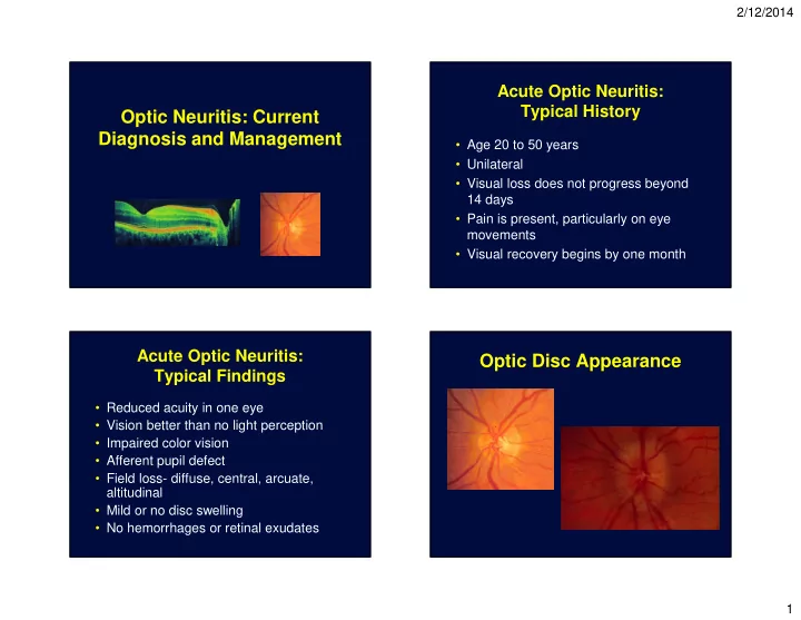
Acute Optic Neuritis: Typical Findings
- Reduced acuity in one eye
- Vision better than no light perception
- Impaired color vision
- Afferent pupil defect
- Field loss- diffuse, central, arcuate,
altitudinal
- Mild or no disc swelling
- No hemorrhages or retinal exudates