
SLIDE 1
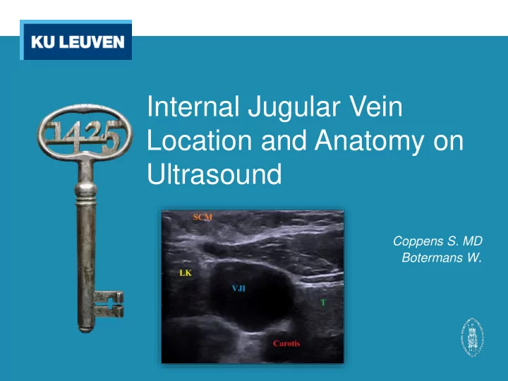
Internal Jugular Vein Location and Anatomy on Ultrasound
Coppens S. MD Botermans W.
Internal Jugular Vein Location and Anatomy on Ultrasound Coppens - - PowerPoint PPT Presentation
Internal Jugular Vein Location and Anatomy on Ultrasound Coppens S. MD Botermans W . Internal Jugular Vein Location and Anatomy on Ultrasound Introduction Methods Results Discussion Conclusion References Introduction
Coppens S. MD Botermans W.
internal jugular vein
specific patient characteristics
Average: Diameter: 13.11 mm Cross-sectional area: 135.52 mm² Side: Left (12.85 mm) vs. right (13.17 mm) p = 0.85 Gender: ♂ (12.48 mm) vs. ♀ (13.73 mm) p = 0.42
Age: ρ* = -0.06 Length: ρ = 0.05 Weight: ρ = 0.11 BMI: ρ = 0.09
* Pearson’s correlation coefficient
Side: Left (83%*) vs. right (29%) RR 2.86; 1.39-5.86** p = 0.03 Gender: (right) ♂ (25%) vs. ♀ (33%) RR 0.75; 0.21-2.66 (left) ♂ (100%) vs. ♀ (67%) RR 1.5; 0.67-3.34
* Percentage of cases with significant overlap ** 95% confidence interval
Age: ρ = 0.04
RR = 4 (not statistically significant)
p = 0.63
RR 2.75 (not statistically significant)
p = 0.02
Literature Study
shorter time window, in general and at first attempt (98% vs. 87%)1,2
↔
performed by residents)
(13.5% vs. 3.9%)1
↔
(attempts performed by residents)
cannulation5
↔
cannulation (not statistically significant)
Literature Study
vessels3
↔
until success in smaller vessels (not
statistically significant)
vein (and is less prone to dilation with Valsalva manoever)4
↔
vein not significant
cannulation5
↔
cannulation (not statistically significant)
Literature Study
to the artery (41,9%) with a variable degree of overlap6
↔
the elderly and when the head is rotated contralaterally, and to a lesser extend on the left side and in men6
↔
and age
significant overlap is present
placement increases success ratio and decreases the risk
anteriorly with a varying degree of overlap
further extend than the right one does
increased risk of complications has been confirmed
1
Brass P, Hellmich M, Kolodziej L, Schick G, Smith AF. Ultrasound guidance versus anatomical landmarks for internal jugular vein catheterization. Cochrane Database of Systematic Reviews 2015, Issue 1. Art. No.: CD006962.
2
Wu SY, Ling Q, Cao LH, Wang J, Xu MX, Zeng WA. Real-time two-dimensional ultrasound guidance for central venous cannulation: a meta-analysis. Anesthesiology. 2013 Feb;118(2):361-75.
3
Mey U, Glasmacher A, Hahn C, Gorschlüter M, Ziske C, Mergelsberg M. Evaluation of an ultrasound-guided technique for central venous access via the internal jugular vein in 493
4
Czyzewska D, Ustymowicz A, Kosel J. Internal jugular veins must be measured before
5
Sulek CA, Blas ML, Lobato EB. A Randomized Study of Left Versus Right Internal Jugular Vein Cannulation in Adults. Journal of Clinical Anesthesia (2000) 12, 142–145.
6
Umaña M, García A, Bustamante L, Castillo JL, Martínez JS. Variations in the anatomical relationship between the common carotid artery and the internal jugular vein: An ultrasonographic study. Colomb Med. 2015; 46(2): 54-59.