Epiretinal 1. How is epiretinal membrane (ERM) best diagnosed? 2. - - PDF document

epiretinal
SMART_READER_LITE
LIVE PREVIEW

Epiretinal 1. How is epiretinal membrane (ERM) best diagnosed? 2. - - PDF document

7/13/18 Financial Disclosure: Nothing to Disclose Learning Objectives Epiretinal 1. How is epiretinal membrane (ERM) best diagnosed? 2. How is ERM differentially diagnosed from central serous Membrane retinopathy and cystoid macular edema?


slide-1
SLIDE 1

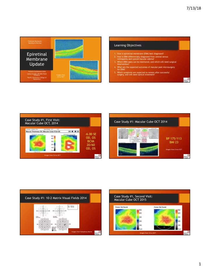
7/13/18 1 Epiretinal Membrane Update

Victoria Conference 2018 James Kundart OD MEd FAAO FCOVD-A Pacific University College of Optometry Financial Disclosure: Nothing to Disclose Images from Cirrus OCT

Learning Objectives

  • 1. How is epiretinal membrane (ERM) best diagnosed?
  • 2. How is ERM differentially diagnosed from central serous

retinopathy and cystoid macular edema?

  • 3. Which ERM cases can be monitored, and which will need surgical

intervention?

  • 4. What are the expected outcomes of macular peel microsurgery

for ERM?

  • 5. Which symptoms are expected to remain after successful

surgery, and will need optical treatment?

Case Study #1, First Visit: Macular Cube OCT , 2014

  • 6.00 SE

OD, OS BCVA 20/60 OD, OS

Images from Cirrus OCT

Case Study #1: Macular Cube OCT 2014 BP 175/113 BMI 23

Images from Cirrus OCT

Case Study #1: 10-2 Matrix Visual Fields 2014

Images from Humphrey Matrix

Case Study #1, Second Visit: Macular Cube OCT 2015

Images from Cirrus OCT

slide-2
SLIDE 2

7/13/18 2

Case Study #1 5-Line Raster OCT 2015

BP 166/84 BMI 23

BCVA 20/50 OD, OS

Images from Cirrus OCT

Case Study #1, Third Visit: Macular Cube OCT Spring 2016

BCVA 20/60 OD and OS

Images from Cirrus OCT

Case Study #1: Macular Cube OCT Spring 2016

Images from Cirrus OCT

Case Study #1: 5-Line Raster Macular OCT Spring 2016

Images from Cirrus OCT

Case Study #1: Macular Cube OCT Summer 2016

Images from Cirrus OCT

Case Study #1: Corneal Topography and 10-2 Matrix Visual Fields 2016

Images from Medmont Topographer and Humphrey Matrix

slide-3
SLIDE 3

7/13/18 3

Case Study #1: Before ERM Peel/Vitrectomy 5-Line Raster OCRT 2017

BCVA 20/70 OD, OS

Images from Cirrus OCT

Case Study #1: Post-Op Peel OS

BCVA 20/50 OD, OS

Case Study #1: Post-Op Peel OS

BCVA 20/50 OD, OS

Case Study #1, Last Visit: 10-2 Matrix Visual Fields and Summary

BP 130/88, BMI 22

Other Causes of ERM: Adult-Offset Coats’ Disease Pre-Op

  • “(a) Color fundus photograph
  • f the right eye of a 38-year-
  • ld man with epiretinal

membrane along supertemporal arcade

  • (b) Ultra-wide-field color

photograph showing exudation and telangiectatic vessels in temporal periphery.”

  • The patient was 20/25 OD with

metamorphopsia OD

https://www.ncbi.nlm.nih.gov/pmc/articles/PMC5678313/

Other Causes of ERM: Adult-Offset Coats’ Disease Post-Op (Peel/Vitrectomy)

https://www.ncbi.nlm.nih.gov/pmc/articles/PMC5678313/

slide-4
SLIDE 4

7/13/18 4

Other Causes of ERM: Eales Disease

https://www.ncbi.nlm.nih.gov/pmc/articles/PMC5859602/

Other Causes of ERM: Eales Disease

https://www.ncbi.nlm.nih.gov/pmc/articles/PMC5859602/

  • (a) Fundus photograph and (b)

fluorescein angiogram of a 22-year-old male with Eales disease Stage 3A and best-corrected visual acuity 0.1 showing fibrovascular proliferation at the disc

  • (c) spectral domain optical coherence

tomography showed cystoid macular edema

  • (j) spectral domain optical coherence

tomography showed epiretinal membrane and macular edema

Epiretinal Membrane Differential Diagnoses: Cystoid Macular Edema (52 YOM and 13 YOF)

http://www.ojoonline.org/article.asp?issn=0974- 620X;year=2012;volume=5;issue=3;spage=187;epage=188;aulast=Garoon

ERM DDx: Cystoid Macular Edema (CME) Using NSAIDs Post-Cataract Surgery

https://www.ncbi.nlm.nih.gov/pmc/articles/PMC5319126/

ERM DDx: Cystoid Macular Edema (CME) Do Steroids Help?

Optical coherence tomography images of cystoid macular edema (CME)

  • Top: Preoperative and postoperative

findings of definite CME

  • Middle: Preoperative and postoperative

findings of probable CME

  • Bottom: Preoperative and postoperative

(cataract surgery) findings of possible CME

https://www.ncbi.nlm.nih.gov/pmc/articles/PMC5636715/

Where ERM Comes From: Latanoprost, CME, and ERM

  • A. Optical coherence tomography of right eye

before latanoprost administration. No pathology is detected.

  • B. Seven months after treatment with

preservative free latanoprost optical coherence tomography in right eye revealed cystoid macular edema with well-defined, intraretinal cystic areas

  • f low reflectivity in the macula with serous retinal

detachment

  • C. Two months after latanoprost discontinuation
  • ptical coherence tomography demonstrated

complete resolution of cystoid macular edema A subtle epiretinal membrane is noted

slide-5
SLIDE 5

7/13/18 5

ERM DDx: Does Latanoprost Cause CME? (after cataract surgery OD)

https://www.ncbi.nlm.nih.gov/pmc/articles/P MC5360021/pdf/13104_2017_Article_2448.pdf

Niacin and CME

http://www.hindawi.com/journals/criopm/2013/713061/

ERM DDx: Microcystic Macular Edema

http://iovs.arvojournals.org/article.aspx?articleid=2190200

ERM DDx: Microcystic Macular Edema (MME)

  • An example of a normal retina compared with

the retina of a patient with MME. (A) A normal retina from a healthy control subject

  • The pseudo-colored surface image is the

infrared surface photo; the vertical stacked gray image an OCT B-scan

  • (B) Optical coherence tomography image

taken from a 76-year-old woman with a 3- year history of AMD, for which she received regular injections with ranibizumab and bevacizumab

  • At time of imaging, her best corrected VA OS

was 0.7 (20/30)

http://iovs.arvojournals.org/article.aspx?articleid=2190200

ERM DDx: Microcystic Macular Edema (MME)

http://iovs.arvojournals.org/article.aspx?articleid=2190200

  • Representative OCT images of patients with MME from the

clinical spectrum are shown

  • The infrared surface photo and OCT image are presented to the

left and the manually segmented INL to the right (A) Microcystic macular edema in the right eye of a 70-year-old male patient with a history of proliferative diabetic retinopathy treated with panretinal photocoagulation (VA OD 0.3 or 20/60) (B) Optical coherence tomography image showing MME 8 months after occlusion of the vena temporalis superior OD in a 66-year-

  • ld female patient (VA OD 0.7 or 20/30)
  • Microcystic macular edema was located in the temporal

superior quadrant of the inner 3-mm EDTRS grid

  • However, INL thickening extended to the periphery. In addition,

hyperreflective spots were observed in all inner retinal layers

ERM DDx: Microcystic Macular Edema

http://iovs.arvojournals.org/article.aspx?articleid=2190200

  • (D) Microcystic macular edema in

a 58-year-old female patient with a newly diagnosed pucker in the right eye (VA OD 0.2 or 20/100)

  • Again, there were multiple

hyperreflective spots in the inner retinal layers

  • (E) Microcystic macular edema 3

months after vitrectomy, in a 71- year-old female patient with a retinal detachment in the left eye (VA OS 0.05 or 20/400)

slide-6
SLIDE 6

7/13/18 6

ERM DDx: Microcystic Macular Edema

http://iovs.arvojournals.org/article.aspx?articleid=2190200

  • Microcystic macular edema in a patient with multiple

sclerosis and a history of optic neuritis and branch retinal vein occlusion in the right eye

  • This 55-year-old female patient was diagnosed with

clinical definite MS in 1992

  • Magnetic resonance imaging showed multiple

periventricular brain lesions, and spinal T2 hyperintense lesions

  • In 2001, she experienced one episode of optic neuritis

OD; coincidentally, an occlusion of the vena temporalis superior was found (VA 0.4 or 20/50)

ERM DDx: Microcystic Macular Edema Propensity for the Nasal/Temporal Quadrants

http://iovs.arvojournals.org/article.aspx?articleid=2190200

  • Most frequently, MME

was observed in patients with ARMD (27.1%) followed by patients with preceding

  • phthalmic surgery

(20.3%) or presence of an epiretinal membrane (18.8%)

Last ERM DDx: Case Study #2

  • A 62-year-old male first reported to our

clinic with a chief concern was a larger image size OS

  • The patient had no history of eye surgery
  • f any sort
  • There was a history of blunt trauma to

the right nasal canthus almost 40 years previously

  • The patient reported that he was

generally healthy with a history of hay fever

https://journals.lww.com/optvissci/Abstract/2018/03000/ Retinal_Nerve_Fiber_Layer_Thickness_in_Various.11.aspx

Case Study #2: Optic Nerve Head OCT

Images from Cirrus OCT

Case Study #2: Optic Nerve and RNFL

Images from Cirrus OCT

Case Study #2: Optic Nerve Head OCT Compare OD to OS

Images from Cirrus OCT

slide-7
SLIDE 7

7/13/18 7

Case Study #2: Optic Nerve OCT

Images from Cirrus OCT

Case Study #2: Retinal Nerve Fiber Layer OCT

Images from Cirrus OCT

Case Study #2: Nerve Fiber Layer Analysis

Images from Cirrus OCT

Case Study #2: Optic Nerve OCT Summary

Images from Cirrus OCT

Case Study #2: Matrix N-30-5 Screener is Unremarkable

  • Why did we run a

frequency-doubling visual field screening?

  • There are two reasons:

both related to cranial nerves

  • Which two nerves were we

checking with his screening field?

Images from Humphrey Matrix

Case Study #2: 24-2 SITA Standard Visual Fields

slide-8
SLIDE 8

7/13/18 8

Case Study #2: Pachymetry with Ant Seg OCT

Images from Cirrus OCT

Case Study #2: Assessment & Initial Plan

http://opticaldiagnostics.com/info/aniseikonia.html

http://cdn.iofferphoto.com/img3/item/568/007/701/o_xalatan- eye-drops-latanoprost-0-005-anti-glaucoma-3932.jpg

Case Study #2, Second Visit: Contact Lens Telescope and New discovery

https://www.flonase.com/allergies/is- flonase-a-nasal-steroid-spray/ http://gogadgetplus.blogspot.com/2014/02/ terminator-telescopic-contact-lens.html

Case Study #2: Macular OCT OD and OS

Images from Cirrus OCT

Case Study #2, Visit 1: Macular OCT OU

Images from Cirrus OCT

Case Study #2, Second Visit: Macular OCT OD

Images from Cirrus OCT

slide-9
SLIDE 9

7/13/18 9

Case Study #2, Visit 2: Macular OCT OS

Images from Cirrus OCT

Case Study #2: Macular OCT OD and OS

Images from Cirrus OCT

Case Study #2: What’s Your Diagnosis OS?

Images from Cirrus OCT

Case Study #2: Macular OCT 5-Line Raster

Images from Cirrus OCT

Case Study #2 Dx: Central Serous Retinopathy (CSR) OS

Images from Cirrus OCT

Case Study #2: Macular OCT OD and OS

Images from Cirrus OCT

slide-10
SLIDE 10

7/13/18 10

Summary: Epiretinal Membrane

  • Epiretinal membrane can be concurrent

and confused with:

  • Eye disease, like Coats’ and Eales
  • Cystoid and Microcystic Macular Edema
  • Central Serous Retinopathy
  • Systemic hypertension leads to some of

these conditions, but not necessarily ERM

  • Aniseikonia is a presenting and residual

symptom, uniquely treated by optometry

  • ERM causes macropsia, magnify the fellow

eye

http://iovs.arvojournals.org/a rticle.aspx?articleid=2458795

Questions? Thank You!

James Kundart OD MEd FAAO FCOVD-A Professor, Pacific University College of Optometry 3D Performance Clinic, Beaverton, Oregon Kundart@pacificu.edu

Additional References

1. Xiao W, Chen X, Yan W, Zhu Z, He M Prevalence and risk factors

  • f epiretinal membranes: a systematic review and meta-analysis of

population-based studies. BMJ Open. 2017 Sep 25;7(9):e014644. https://www.ncbi.nlm.nih.gov/pmc/articles/PMC5623383/ 2. Shen Z, Duan X, Wang F , Wang N, Peng Y , Liu DT , Peng X, Li S, Liang Y . Prevalence and risk factors of posterior vitreous detachment in a Chinese adult population: the Handan eye study. BMC Ophthalmol. 2013 Jul 16;13(1):33. https://www.ncbi.nlm.nih.gov/pmc/articles/PMC3726418/ 3. Daruich A, Matet A, Dirani A, Bousquet E, Zhao M, Farman N, Jaisser F , Behar- Cohen F . Central serous chorioretinopathy: Recent findings and new physiopathology hypothesis. Prog Retin Eye Res. 2015 Sep;48:82-118. https://www.sciencedirect.com/science/article/pii/S1350946215000336?via% 3Dihub