
SLIDE 1
Sandia is a multi-program laboratory operated by Sandia Corporation, a Lockheed Martin Company, for the United States Department of Energy under contract DE-AC04-94AL85000.
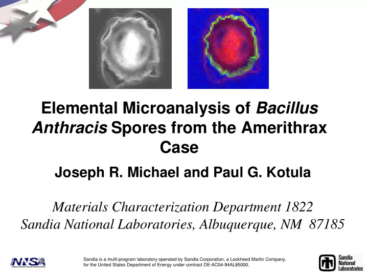
Elemental Microanalysis of Bacillus Anthracis Spores from the - - PowerPoint PPT Presentation
Elemental Microanalysis of Bacillus Anthracis Spores from the Amerithrax Case Joseph R. Michael and Paul G. Kotula Materials Characterization Department 1822 Sandia National Laboratories, Albuquerque, NM 87185 Sandia is a multi-program
Sandia is a multi-program laboratory operated by Sandia Corporation, a Lockheed Martin Company, for the United States Department of Energy under contract DE-AC04-94AL85000.
Volume excited ~ 1 μm3 Volume excited ~ 10-8 μm3 100 nm
1 nm 1 nm
In this study we make use of the characteristic x-rays generated by the electron/sample interactions.
1 2 3
2 4 6 8 10 0.1 0.2 0.3 0.4 0.5
Ti
x y energy pixel
energy
W-M
W-L
Co Ni
Co-L Ni-L
Sn-L
5 10 15
Focused electron probe Distribution of elemental x-ray signals Red = C Red = C-
support Green = alumina Green = alumina Blue = Blue = FeCo FeCo Cyan = Ca Cyan = Ca-
S-
Si-
O Black = shadowed support Black = shadowed support
Keenan, M. R., and Kotula, P. G.,(2003) Apparatus and System for Multivariate Spectral Analysis., US Patent #6584413. (filing date June 1, 2001). Keenan, M. R., and Kotula, P. G.,(2004) Method of Multivariate Spectral Analysis., US Patent #6675106. (filing date June 1, 2001) Kotula, P. G., Keenan, M. R., Michael, J. R. (2003), “Automated Analysis of SEM X-ray Spectral Images: A Powerful New Microanalysis Tool, Microscopy and Microanalysis; Feb. 2003; vol.9, no.1, pp.1-17.
Sample fixation/ inactivation Gamma irradiation (4Mrad) or 1 %Osmium tetroxide (1 hour) or Glutaraldehyde (96 hours) Rinse in Millonig’s buffer Dry powder sample Dry powder sample SEM Access sample and dust on stub in disposable glove bag Image sample either uncoated (variable pressure SEM) or after conducive coating Access sample and mount on stub in disposable glove bag Mount sample in FIB and ion mill thin sample from spore(s) Move thin sample to carbon film
Dehydration (30% ethanol) 50% ethanol 70% ethanol 90% ethanol 100% ethanol 100% propylene
Embedding 1:1 propylene oxide:resin 100% resin Place in mold with fresh resin and cure (oven)
Section and collect
Stain Uranyl Acetate Lead citrate ( S ) T E M (S)TEM Performed at USAMRIID or NBFAC Performed at Sandia National Laboratories Dry powder sample (S)TEM Access sample and dust on TEM grid in disposable glove bag
100 0 200 0 300 0 400 0 500 0 600 0 700 0 1 2 3 4 5
Counts Energy (kV)
1.00 2.00 3.00 4.00 5.00 0.05 0.1 0.15 0.2 0.25 0.3 0.35 0.4 0.45 0.5 10 20 30 40 1.00 2.00 3.00 4.00 5.00 0.1 0.2 0.3 0.4 0.5 0.6 0.7 0.8 0.9 5 10 15 20
keV
1.00 2.00 3.00 4.00 5.00 0.02 0.04 0.06 0.08 0.1 0.12
P Cl K Ca
10 20 30
keV
1.00 2.00 3.00 4.00 5.00 0.02 0.04 0.06 0.08 0.1 0.12 0.14 0.16 10 20 30
P Mg Cl K Na O
Ca-P
500 1000 1500 2000 2500 1 2 3 4 5 6
500 1000 1500 2000 2500 3000 3500 1 2 3 4 5 6
C O Ca Si P S NaMg
C O Ca Si S NaMg P
20 kV 15 kV 5 kV
Intensity (counts) 1 2 4 3 Energy (keV)
Ca P Si Mg S S
Lower voltages produce more surface elemental information. Very small amount of Si detected at 5 kV therefore Si is locate away from the spore surface.
Si = 1.2 - 2.3 wt% ±50% Ca =3.1 - 6.5 wt% ±50% Si = 1.2 - 1.5 wt% ±50% Ca =2.7 – 3.1 wt% ±50%
5 kV= 300 nm 15 kV = 2100 nm 20 kV= 3300 nm
*L. F. Carey, D. C. St. Amant and M. A. Guelta, Production of Bacillus spores as a simulant for biological warfare agents, Edgewood Chemical Biological Center,ECBE-TR-372, April 2004.
SEM Image of Leahy material
2.00 4.00 6.00 0.2 0.4 0.6 0.8 1 1.2 2.00 4.00 6.00 0.02 0.04 0.06 0.08 0.1 5 10 15 5 10 15
C
keV keV
Ca P Si S O Na Mg
Spectral Image components of Leahy material
Volume excited ~ 1 μm3 Volume excited ~ 10-8 μm3 100 nm
1 nm 1 nm
In this study we make use of the characteristic x-rays generated by the electron/sample interactions.
Sample fixation/ inactivation Gamma irradiation (4Mrad) or 1 %Osmium tetroxide (1 hour) or Glutaraldehyde (96 hours) Rinse in Millonig’s buffer Dry powder sample Dry powder sample SEM Access sample and dust on stub in disposable glove bag Image sample either uncoated (variable pressure SEM) or after conducive coating Access sample and mount on stub in disposable glove bag Mount sample in FIB and ion mill thin sample from spore(s) Move thin sample to carbon film
Dehydration (30% ethanol) 50% ethanol 70% ethanol 90% ethanol 100% ethanol 100% propylene
Embedding 1:1 propylene oxide:resin 100% resin Place in mold with fresh resin and cure (oven)
Section and collect
Stain Uranyl Acetate Lead citrate ( S ) T E M (S)TEM Performed at USAMRIID or NBFAC Performed at Sandia National Laboratories Dry powder sample (S)TEM Access sample and dust on TEM grid in disposable glove bag
See: L. N. Brewer, J. A. Ohlhausen, P. G. Kotula and J. R. Michael, Forensic imaging of bioagents by X-ray and TOF- SIMS hyperspectral imaging”, Forensic Science International, vol. 179, 2008, 98-106.
keV
2.00 4.00 6.00 2 4 6 8 200 600 1000
Si O Cl
5.00 10.00 0.05 0.1 0.15 0.2 5.00 10.00 0.05 0.1 0.15 0.2 0.25 0.3 20 40 60 80 100 20 40 60 80
5.00 10.00 0.05 0.1 0.15 0.2 0.25 0.3 5.00 10.00 0.05 0.1 0.15 0.2 20 40 10 20 30 40
Si O Pb U Ni Pb Os U Ni Os
Carbon in support media
Fe
Ion image of TEM specimen SEM of TEM specimen ready to be extracted <100nm thick STEM sample Region of TEM sample
STEM image
2.00 4.00 6.00 0.1 0.2 0.3 0.4 2.00 4.00 6.00 0.05 0.1 0.15 0.2 20 40 60 10 20 30
Si O Fe Ca K P Mg Na O
3.00 4.00 5.00 6.00 7.00 0.005 0.01 0.015 0.02
Sn
Sn Fe
0.1 0.2 0.3 0.4 0.5 0.6 0.7 0.8 0.9 1
10µm
10 20 30 40 50 60 70 80 90 100 110 120 130 140 150 160 170 180 190 200 5 10 15
Mass / Charge 110 120 130
0.0 0.1 0.2 0.3 0.4 0.5
Si K Ga Fe SiOH Ca Sn Sn
2.00 4.00 6.00 0.1 0.2 0.3 0.4 0.5 2.00 4.00 6.00 0.1 0.2 0.3 0.4 0.5 2.00 4.00 6.00 0.2 0.4 0.6 0.8 1 1.2 1.4 10 20 10 20 30 20 40 60 80
Sn Si O C C N S O Ca
2.00 4.00 0.02 0.04 0.06 0.08 0.1
Si P S O C Al Fe Sn
P C O K Na Mg
ADF STEM image
2.00 4.00 6.00 0.1 0.2 0.3 0.4 5 10 15 2.00 4.00 6.00 0.1 0.2 0.3 0.4 0.5 0.6 0.7 2.00 4.00 6.00 0.05 0.1 0.15 0.2 0.25 0.3 0.35 10 20 30 5 10 15 20 25
Ca P K O C S C O Si O C S Sn
Fe Na Mg
2.00 4.00 0.02 0.04 0.06 0.08 0.1 1.00 2.00 3.00 0.05 0.1 0.15 0.2 10 20 30 20 40
O Si keV P + Os Cl Ca
2.00 4.00 6.00 0.1 0.2 0.3 0.4 0.5 20 40 60 80
Sn Si O C keV Fe
2.00 4.00 6.00 0.1 0.2 0.3 0.4 20 40 60
Si O Fe Sn
5.00 10.00 0.05 0.1 0.15 0.2 0.25 0.3 20 40 60 80
Fe Si O keV
Daschle Material New York Post Material Leahy Material
Spore coats on Leahy and New York Post samples are indistinguishable (both contain Si, Fe and Sn. Daschle appears the same (Si and Fe present, Sn is obscured by other elements in stain). Material from the Daschle letter was not made available for FIB sectioning.
X Number of spores with a particular chemical feature n Total number of spores analyzed
0.1 0.2 0.3 0.4 0.5 0.6 0.7 0.8 0.9 1
500 nm
1 2 3 4 5 6 7 8 9 10
Energy (keV)
Red= Si, Green = Ca-P, Blue, Cl-S
Background Coat Cortex Core
*Johnstone, K. et al., Location of metal ions on Bacillus megaterium spores by
high-resolution electron probe x-ray microanalysis, FEMS microbiology Letters,
Modifed CCY medium containing: MgCl26H2O, MnCl2 4H2O, FeCl36H2O, ZnCl2, CaCl26H2O, KH2PO4, K2HPO4, glutamine, acid casein hydrolysate, enzymatic casein hydrolysate, enzymatic yeast extract and glycerol.
Bact., July 1980, p. 481- 491
0.1 0.2 0.3 0.4 0.5 0.6
1 2 3 4 5 6 7 8 9 10
Mg Si P S Ca Mn Cu from grid
Author’s noted: “considerable variation in Si content both within and between different spore preparations,… unlikely to be due entirely to contamination.”