
SLIDE 1
7/3/2011 1
Disorders of Blood Cells & Blood Coagulation g
HIHIM 409
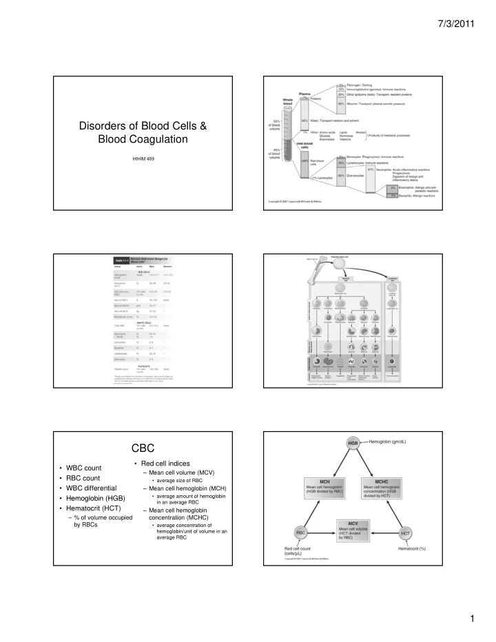
CBC
- WBC count
- RBC count
- WBC differential
- Hemoglobin (HGB)
- Red cell indices
– Mean cell volume (MCV)
- average size of RBC
– Mean cell hemoglobin (MCH)
- average amount of hemoglobin
- Hemoglobin (HGB)
- Hematocrit (HCT)
– % of volume occupied by RBCs
g g in an average RBC
– Mean cell hemoglobin concentration (MCHC)
- average concentration of
hemoglobin/unit of volume in an average RBC